. 2005 Dec 15;392(Pt 3):607-14.
doi: 10.1042/BJ20050671.

Specific role for p85/p110beta in GTP-binding-protein-mediated activation of Akt

Affiliations

Specific role for p85/p110beta in GTP-binding-protein-mediated activation of Akt

Hiroshi Kubo et al. Biochem J. .

Abstract

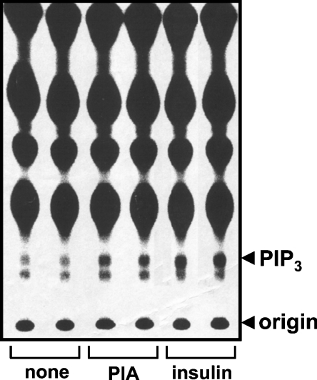
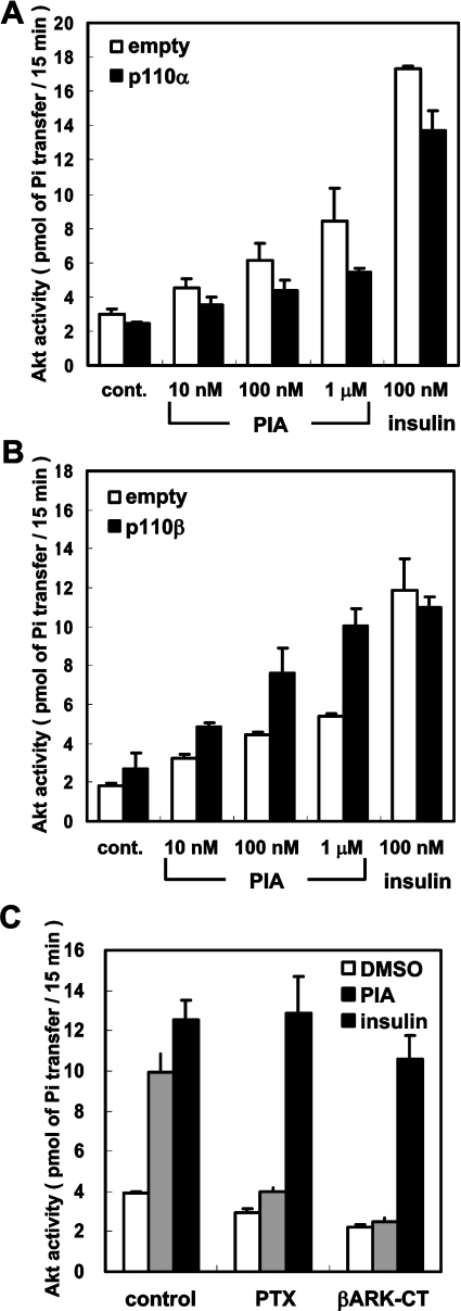
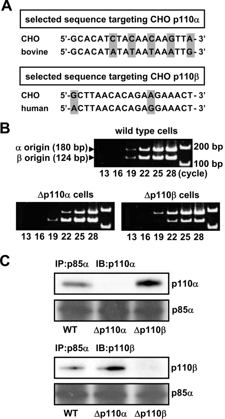
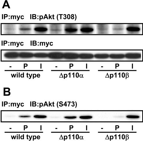
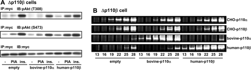
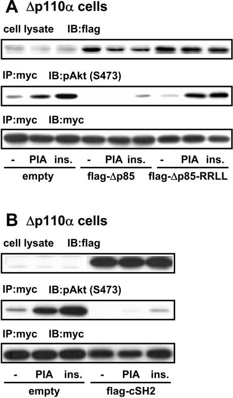
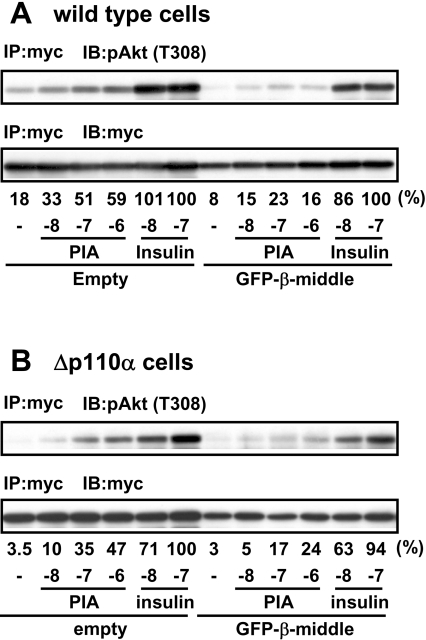
We prepared CHO (Chinese hamster ovary) cells expressing both IR (insulin receptor) and A1R (A1 adenosine receptor). Treatment of the cells with insulin or PIA [N6-(2-phenylisopropyl)adenosine], a specific A(1)R agonist increased Akt activity in the cells in a PI3K- (phosphoinositide 3-kinase) dependent manner. Transfection of p110beta into the cells augmented the action of PIA with little effect on insulin. Introduction of a pH1 vector producing shRNA (short hairpin RNA) that targets p110beta abolished PIA-induced Akt activation. By contrast, an shRNA probe targeting p110alpha did not impair the effects of PIA. The effect of PIA in p110alpha-deficient cells was attenuated effectively by both Deltap85 and betaARK-CT (beta-adrenergic receptor kinase-C-terminal peptide). A Deltap85-derived protein possessing point mutations in its two SH2 domains did not impair PIA action. These results suggest that tyrosine-phosphorylated proteins and Gbetagamma (betagamma subunits of GTP-binding protein) are necessary for the specific function of p110beta in intact cells. The p110beta-middle (middle part of p110beta) may play an important role in signal reception from GPCRs (GTP-binding-protein-coupled receptor), because transfection of the middle part impaired PIA sensitivity.


Figures

Figure 1
Figure 1. Effects of PIA and insulin on PtdIns(3,4,5)P3 production in CHO-IR-A1R cells
CHO-IR-A1R cells, labelled with 32Pi, were stimulated with 1 μM PIA or 100 nM insulin for 5 min. Phospholipids were extracted and separated on oxalate-impregnated TLC plates. An autoradiogram of a TLC plate is shown. PIP3, PtdIns(3,4,5)P3.
Figure 2
Figure 2. Effects of p110β expression on PIA-induced Akt activation
CHO-IR-A1R cells transfected with Myc–Akt were stimulated with PIA or insulin for 4 min. The cell lysate was mixed with anti-Myc antibody and the immune complex was assayed for protein kinase activity with Crosstide as the substrate. Each bar represents the mean±S.D. of triplicate determinations. In (A) p110α or control empty vector was co-transfected with Myc–Akt. In (B) p110β or control empty vector was co-transfected with Myc–Akt. In (C) p110β together with βARK-CT or control vector was co-transfected with Myc–Akt. Treatment with pertussis toxin (PTX) was performed by incubating the cells with 10 ng/ml of the toxin for 16 h before stimulation.
Figure 3
Figure 3. Preparation of Δp110α and Δp110β cells
(A) The panel shows the sense sequences of the CHO versions of p110α and p110β genes selected as targets of the siRNA gene-silencing technique. The corresponding sequences of bovine p110α and human p110β are also shown. (B) CHO-IR-A1R cells were transfected with the pH1 expression vector and producing shRNA targeting the sequences of CHO-p110α and CHO-p110β using LipofectAMINE Plus™, cells resistant to 8 μg/ml puromycin were selected. Total RNA from the cells was analysed for the mRNAs of p110α and p110β by RT-PCR using gene-specific primers. (C) The lysate from Δp110α or Δp110β cells was mixed with the anti-serum against p85 and the immune complex was subjected to Western blotting using the specific antibody against p110α or p110β. IB, immunoblot; IP, immunoprecipitation.
Figure 4
Figure 4. Effect of p110β knockdown on PIA-induced Akt activation
(A) The Δp110α or Δp110β cells transfected with Myc–Akt were stimulated with 1 μM PIA (P) or 0.1 μM insulin (I). The cell lysate was subjected to immunoprecipitation (IP) with anti-Myc antibody followed by Western blotting with anti-Myc and anti-pAkt (pT308) antibodies (IB). (B) The blot in (A) was reprobed with anti-pAkt (pS473) antibody.
Figure 5
Figure 5. Recovery of PIA action by transfection of shRNA-resistant p110β
(A) The Δp110β cells were transfected with Myc–Akt, together with bovine version of p110α or human version of p110β. The cells were then stimulated with 1 μM PIA or 0.1 μM insulin (ins.). The cell lysate was subjected to immunoprecipitation with anti-Myc antibody (IP) followed by Western blotting with anti-pAkt (pT308), anti-pAkt (pS473) and anti-Myc antibodies (IB). (B) Total RNA from the cells was analysed for the mRNAs of CHO-p110α, CHO-p110β, bovine p110α and human p110β by RT-PCR using gene-specific primers.
Figure 6
Figure 6. Effects of Δp85 on the action of PIA in p110α-deficient cells
Δp110α cells were transfected with Myc–Akt, together with FLAG-Δp85 (in A), FLAG-Δp85-R358L/R649L (FLAG-Δp85-RRLL in A), FLAG-tagged C-SH2 domain of p85 (FLAG-cSH2 in B) or empty vector. After stimulation of the cells with 1 μM PIA or 0.1 μM insulin, the cell lysate was prepared and subjected to immunoprecipitation with anti-Myc antibody (IP). The immune complex was analysed by Western blotting with anti-FLAG, anti-pAkt (pS473) and anti-Myc antibodies (IB).
Figure 7
Figure 7. Effect of p110β-middle protein on the action of PIA
Wild-type CHO-IR-A1R cells (A) or Δp110α cells (B) were transfected with Myc–Akt, together with GFP-p110β-middle or empty vector and then stimulated with PIA or insulin. The cell lysate was subjected to immunoprecipitation with anti-Myc antibody (IP) followed by Western blotting with anti-pAkt (pT308) and anti-Myc antibodies (IB). The blots were analysed by densitometer and the density of pAkt was normalised to that of Myc. The relative values are presented just below the autoradiogram, as percentage of 0.1 μM insulin in vector-control cells; the values below this are the concentrations of PIA or insulin (mole/l), represented by logarithm unit {log[M]}.

Similar articles

Cited by

References

    1. Shepherd P. R., Withers D. J., Siddle K. Phosphoinositide 3-kinase: the key switch mechanism in insulin signalling. Biochem. J. 1998;333:471–490. - PMC - PubMed
    1. Backer J. M., Myers M. G., Jr, Shoelson S. E., Chin D. J., Sun X. J., Miralpeix M., Hu P., Margolis B., Skolnik E. Y., Schlessinger J., White M. F. Phosphatidylinositol 3′-kinase is activated by association with IRS-1 during insulin stimulation. EMBO J. 1992;11:3469–3479. - PMC - PubMed
    1. Carpenter C. L., Auger K. R., Chanudhuri M., Yoakim M., Schaffhausen B., Shoelson S., Cantley L. C. Phosphoinositide 3-kinase is activated by phosphopeptides that bind to the SH2 domains of the 85-kDa subunit. J. Biol. Chem. 1993;268:9478–9483. - PubMed
    1. Yu J., Zhang Y., McIlroy J., Rordorf Nikolic T., Orr G. A., Backer J. M. Regulation of the p85/p110 phosphatidylinositol 3′-kinase: stabilization and inhibition of the p110α catalytic subunit by the p85 regulatory subunit. Mol. Cell. Biol. 1998;18:1379–1387. - PMC - PubMed
    1. Brock C., Schaefer M., Reusch H. P., Czupalla C., Michalke M., Spicher K., Schultz G., Nurnberg B. Roles of Gβγ in membrane recruitment and activation of p110γ/p101 phosphoinositide 3-kinase γ. J. Cell Biol. 2003;160:89–99. - PMC - PubMed