. 2005 Aug 9:4:9.
doi: 10.1186/1475-2883-4-9.

Studies on Acanthocheilonema viteae cystatin: genomic organization, promoter studies and expression in Caenorhabditis elegans

Affiliations

Studies on Acanthocheilonema viteae cystatin: genomic organization, promoter studies and expression in Caenorhabditis elegans

Smitha Pillai et al. Filaria J. .

Abstract

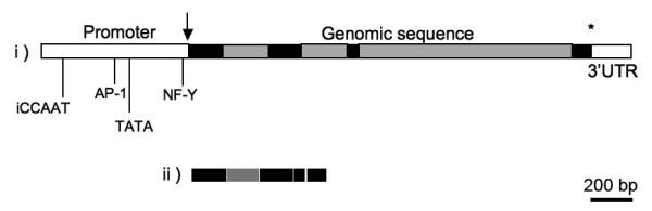
Cystatins are reversible, tightly binding inhibitors of cysteine proteases. Filarial cystatins have been ascribed immunomodulatory properties and have been implicated in protective immunity. To continue exploration of this potential, here we have determined the sequence, structure and genomic organization of the cystatin gene locus of A. viteae. The gene is composed of 4 exons separated by 3 introns and spans approximately 2 kb of genomic DNA. The upstream genomic sequence contains transcriptional factor binding sites such as AP-1 and NF-Y, an inverted CCAAT sequence, and a TATA box. To investigate sites of cystatin expression, Caenorhabditis elegans worms were transformed by microinjection with the putative promoter region and the first exon of the A. viteae cystatin gene fused to the reporter GFP. In transgenic worms fluorescence was observed in the pharyngeal and rectal gland cells suggesting that cystatin is secreted. Additionally, A. viteae cystatin was expressed in C. elegans to explore its potential as an expression system for filarial genes.


Figures

Figure 1
Figure 1
The nucleotide sequence of Acanthocheilonema viteae cystatin gene showing the 5' upstream genomic sequence, four exons interrupted by three introns and the 3' UTR. The putative TATA box and transcription factor binding sites (CCAAT box, two AP-1 binding sites (CCAAT) and one NF-Y binding site (TGAGTCA)) are boxed. The sequence highlighted in gray shading corresponds to the 5' upstream genomic sequence and 3' UTR. Exons are in capital letters. The start ATG and the stop TGA are italizised.
Figure 2
Figure 2
Expression pattern of the Av17promoter::GFP construct in transgenic Caenorhabditis elegans. (A) Fluorescence staining in the pharynx of a worm transformed with the Av17promoter::GFP fusion construct. The pharynx contains two classes of gland cells, g1 (two cells) and g2 (two cells) in the second bulb of the pharynx. The g1 cells extend three cuticle-lined ducts anteriorly within the narrow pharyngeal nerve cords, two of which pass through the isthmus and empty into the pharyngeal lumen near the first bulb and the dorsal g1 duct empties near the anterior end of the pharynx. The g2 cells extend shorter ducts, which empty into the lumen of the second bulb. (C) Fluorescence staining in the rectal gland cells (rectD, rectVL, rectVR) in the posterior of the same worm as in (A). These cells connect to the intestinal lumen just posterior to the rectal valve. (B) and (D) Bright field microscopy of (A) and (C), respectively.
Figure 3
Figure 3
(A) Single worm PCR of transgenic worms, 49cAv17 and 49gAv17, containing the cDNA (lane 1) and genomic sequence (lane 2) of A. viteae cystatin respectively. (B) RT-PCR analysis of transcripts from transgenic worms, 49cAv17 and 49gAv17, containing the cDNA (lane 2) and genomic sequence (lane 3) of A. viteae cystatin respectively. Negative control with wild type C. elegans in lane 4 and marker in lane 1. (C) Western Blot with anti-His antibodies. Lane 1: Transgenic C. elegans expressing Av17 constitutively (103cAv173') ; Lane 2: Transgenic C. elegans (49cAv17) expressing Av17 induced by heat shock ; Lane 3: pha-1 control worms
Figure 4
Figure 4
i) Schematic representation of the genomic structure of Av17 (black and grey boxes represent the exons and the introns respectively). ii) intron splicing of genomic sequence of Av17 C. elegans (103cAv173').

Similar articles

References

    1. Maizels RM, Lawrence RA. Immunological tolerance: The key feature in human filariasis? Parasitol Today. 1991;7:271–276. doi: 10.1016/0169-4758(91)90093-4. - DOI - PubMed
    1. Nicklin MJ, Barrett AJ. Inhibition of cysteine proteinases and dipeptidyl peptidase I by egg-white cystatin. Biochem J. 1984;223:245–253. - PMC - PubMed
    1. Lustigman S, Brotman B, Huima T, Prince AM. Characterization of an Onchocerca volvulus cDNA clone encoding a genus specific antigen present in infective larvae and adult worms. Mol Biochem Parasitol. 1991;45:65–75. doi: 10.1016/0166-6851(91)90028-5. - DOI - PubMed
    1. Hartmann S, Kyewski B, Sonnenburg B, Lucius R. A filarial cysteine protease inhibitor down-regulates T cell proliferation and enhances interleukin-10 production. Eur J Immunol. 1997;27:2253–2260. - PubMed
    1. Maizels RM, Gomez-Escobar N, Gregory WF, Murray J, Zang X. Immune evasion genes from filarial nematodes. Int J Parasitol. 2001;31:889–898. doi: 10.1016/S0020-7519(01)00213-2. - DOI - PubMed

LinkOut - more resources