. 2005 Aug 10:6:106.
doi: 10.1186/1471-2164-6-106.

Cross genome phylogenetic analysis of human and Drosophila G protein-coupled receptors: application to functional annotation of orphan receptors

Affiliations

Cross genome phylogenetic analysis of human and Drosophila G protein-coupled receptors: application to functional annotation of orphan receptors

Raghu Prasad Rao Metpally et al. BMC Genomics. .

Abstract

Background: The cell-membrane G-protein coupled receptors (GPCRs) are one of the largest known superfamilies and are the main focus of intense pharmaceutical research due to their key role in cell physiology and disease. A large number of putative GPCRs are 'orphans' with no identified natural ligands. The first step in understanding the function of orphan GPCRs is to identify their ligands. Phylogenetic clustering methods were used to elucidate the chemical nature of receptor ligands, which led to the identification of natural ligands for many orphan receptors. We have clustered human and Drosophila receptors with known ligands and orphans through cross genome phylogenetic analysis and hypothesized higher relationship of co-clustered members that would ease ligand identification, as related receptors share ligands with similar structure or class.

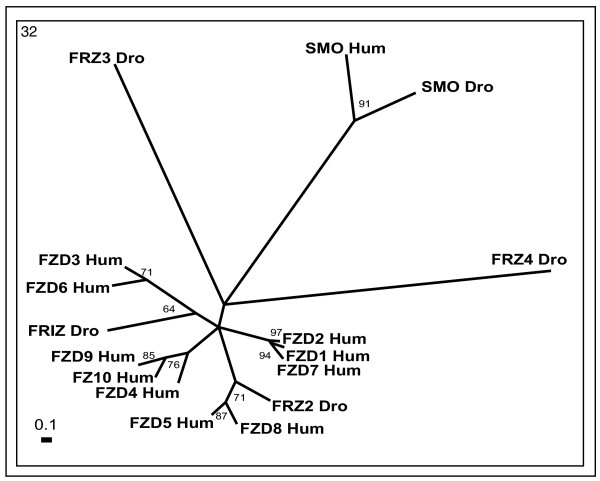
Results: Cross-genome phylogenetic analyses were performed to identify eight major groups of GPCRs dividing them into 32 clusters of 371 human and 113 Drosophila proteins (excluding olfactory, taste and gustatory receptors) and reveal unexpected levels of evolutionary conservation across human and Drosophila GPCRs. We also observe that members of human chemokine receptors, involved in immune response, and most of nucleotide-lipid receptors (except opsins) do not have counterparts in Drosophila. Similarly, a group of Drosophila GPCRs (methuselah receptors), associated in aging, is not present in humans.

Conclusion: Our analysis suggests ligand class association to 52 unknown Drosophila receptors and 95 unknown human GPCRs. A higher level of phylogenetic organization was revealed in which clusters with common domain architecture or cellular localization or ligand structure or chemistry or a shared function are evident across human and Drosophila genomes. Such analyses will prove valuable for identifying the natural ligands of Drosophila and human orphan receptors that can lead to a better understanding of physiological and pathological roles of these receptors.


Figures

Figure 1
Figure 1
Phylogenetic trees of peptide receptors (clusters 1–11). Trees were inferred as described in Methods (using TREE-PUZZLE 5.1 corrected using JTT substitution frequency matrix. Quartet-puzzling support percentage values from 10,000 puzzling steps are shown). Out-group not showed in the figure. The scale bars indicate a maximum likelihood branch length of 0.1 inferred substitutions per site. Orphan receptors are shown in bold letters. Cluster numbers are marked in the top left corner.
Figure 2
Figure 2
Representative multiple sequence alignment of GPCR clusters. GPCR sequences of ET1R_Hum, ETAR_Hum, ETBR_Hum, ETB2_Hum, GRPR_Hum, NMBR_Hum, BRS3_Hum, GP37_Hum, Q8TDV0_Hum, Q9V858_Dro and Q9V9K3_Dro belonging to cluster 4 were aligned with ClustalX. Sequence region comprising of TMH-1 to TMH-7 alone were considered for the analysis (Alignment was modified by deleting the extremely variable amino termini upstream of the first transmembrane helix and carboxyl termini downstream of the seventh transmembrane helix). Identical amino-acid residues in all aligned sequences are shaded in black and similar residues in gray and consensus residues are indicated below. Transmembrane helices (TMH) identified by the HMMTOP program are indicated.
Figure 3
Figure 3
Phylogenetic trees of chemokine receptors (clusters 12 and 13). The mode of deriving phylogenetic trees is as described in Methods and indications are as in Figure 2.
Figure 4
Figure 4
Phylogenetic trees of nucleotide and lipid receptors (clusters 14–19). The mode of deriving phylogenetic trees is as described in Methods and indications are as in Figure 2.
Figure 5
Figure 5
Phylogenetic trees of biogenic amine receptors (clusters 20–24). The mode of deriving phylogenetic trees is as described in Methods and indications are as in the Figure 2 except for the cluster 22, where scale bar indicates a maximum likelihood branch length of 1.0 inferred substitutions per site.
Figure 6
Figure 6
Phylogenetic trees of class B (secretin) receptors (clusters 25 and 26). The mode of deriving phylogenetic trees is as described in Methods and indications are as in Figure 2.
Figure 7
Figure 7
Phylogenetic tree of cell adhesion receptors (cluster 27). The mode of deriving phylogenetic tree is as described in Methods and indications are as in Figure 2.
Figure 8
Figure 8
Phylogenetic trees of class C (glutamate) receptors (clusters 28–31). The mode of deriving phylogenetic trees is as described in Methods and indications are as in Figure 2.
Figure 9
Figure 9
Phylogenetic tree of frizzled/smoothened receptors (cluster 32). The mode of deriving phylogenetic tree is as described in Methods and indications are as in Figure 2.

Similar articles

Cited by

References

    1. Christopoulos A. Allosteric binding sites on cell-surface receptors: novel targets for drug discovery. Nat Rev Drug Discou. 2002;1:198–210. doi: 10.1038/nrd746. - DOI - PubMed
    1. Palczewski K, Kumasaka T, Hori T, Behnke CA, Motoshima H, Fox BA, Le Trong I, Teller DC, Okada T, Stenkamp RE, Yamamoto M, Miyano M. Crystal structure of rhodopsin: A G protein-coupled receptor. Science. 2000;289:739–745. doi: 10.1126/science.289.5480.739. - DOI - PubMed
    1. Libert F, Schiffmann SN, Lefort A, Parmentier M, Gerard C, Dumont JE, Vanderhaeghen JJ, Vassart G. The orphan receptor cDNA RDC7 encodes an A1 adenosine receptor. Embo J. 1991;10:1677–1682. - PMC - PubMed
    1. Pyne S, Pyne NJ. Sphingosine 1-phosphate signalling in mammalian cells. Biochem J. 2000;349:385–402. doi: 10.1042/0264-6021:3490385. - DOI - PMC - PubMed
    1. Marchese A, George SR, Kolakowski LFJ, Lynch KR, O'Dowd BF. Novel GPCRs and their endogenous ligands: expanding the boundaries of physiology and pharmacology. Trends Pharmacol Sci. 1999;20:370–375. doi: 10.1016/S0165-6147(99)01366-8. - DOI - PubMed

Publication types

MeSH terms

LinkOut - more resources