. 2005 Dec 1;106(12):3824-30.
doi: 10.1182/blood-2005-05-2150. Epub 2005 Aug 9.

The alphaC domains of fibrinogen affect the structure of the fibrin clot, its physical properties, and its susceptibility to fibrinolysis

Affiliations

The alphaC domains of fibrinogen affect the structure of the fibrin clot, its physical properties, and its susceptibility to fibrinolysis

Jean-Philippe Collet et al. Blood. .

Abstract

The functions of the alphaC domains of fibrinogen in clotting and fibrinolysis, which have long been enigmatic, were determined using recombinant fibrinogen truncated at Aalpha chain residue 251. Scanning electron microscopy and confocal microscopy revealed that the fibers of alpha251 clots were thinner and denser, with more branch points than fibers of control clots. Consistent with these results, the permeability of alpha251 clots was nearly half that of control clots. Together, these results suggest that in normal clot formation, the alphaC domains enhance lateral aggregation to produce thicker fibers. The viscoelastic properties of alpha251 fibrin clots differed markedly from control clots; alpha251 clots were much less stiff and showed more plastic deformation, indicating that interactions between the alphaC domains in normal clots play a major role in determining the clot's mechanical properties. Comparing factor XIIIa cross-linked alpha251 and control clots showed that gamma chain cross-linking had a significant effect on clot stiffness. Plasmin-catalyzed lysis of alpha251 clots, monitored with both macroscopic and microscopic methods, was faster than lysis of control clots. In conclusion, these studies provide the first definitive evidence that the alphaC domains play an important role in determining the structure and biophysical properties of clots and their susceptibility to fibrinolysis.


Figures

Figure 1.
Figure 1.
Factor XIIIa–induced cross-linking of Aα251 and control fibrinogen. Factor XIII and thrombin were mixed, left on ice for less than 10 minutes, and added to fibrinogen at ambient temperature. The samples with final concentrations of 1 IU/mL factor XIII, 1 IU/mL thrombin, and 1.2 μM fibrinogen were incubated for the indicated times (minutes). The reactions were stopped by addition of SDS (1% final concentration) and incubation in boiling water for 5 minutes; samples were analyzed on 12% SDS-PAGE run under reducing conditions. The 0 time point samples contained only fibrinogen. Positions of molecular weight markers are indicated on the right; positions of bands corresponding to individual fibrin chains are indicated on the left. (A) Control fibrin. (B) α251 fibrin.
Figure 2.
Figure 2.
Images of α251 and control fibrin clots from laser scanning confocal microscopy. Thrombin at a final concentration of 0.9 IU/mL was added to recombinant control or Aα251 fibrinogen at a 4.4-μM final concentration in 20 mM HEPES, pH 7.4, 0.15 M NaCl, 5 mM CaCl2. Images of 3-dimensional reconstructions of optical sections of control (A, B) and α251 (C, D) fibrin networks obtained by laser scanning confocal microscopy. Original magnifications are as follows: panels A and C: 48 × 48 × 48 μm3; panels B and D: 4 × 24 × 24 μm3.
Figure 3.
Figure 3.
Scanning electron microscopy of α251 and control fibrin. Thrombin at a final concentration of 0.9 IU/mL was added to recombinant control or Aα251 fibrinogen at a 4.4-μM final concentration in 20 mM HEPES, pH 7.4, 0.15 M NaCl, 5 mM CaCl2. (A) Fibrin made from control fibrinogen. (B) Fibrin made from control fibrinogen in the presence of 1 IU/mL factor XIIIa. (C) Fibrin clot made from Aα251 fibrinogen. (D) Fibrin clot made from Aα251 fibrinogen in the presence of 1 IU/mL factor XIIIa. Magnification bar equals 1000 nm.
Figure 4.
Figure 4.
Fibrinolysis of α251 and control fibrin clots as visualized by laser scanning confocal microscopy. Series of micrographs showing the dynamic lysis of control (top panels) and α251 (bottom panels) fibrin clots by rtPA (6 nM) and Glu-plasminogen (2.5 μg/mL); clots were prepared as described in Figure 2. Lysis-front motion was visualized by scanning this region of the clot every 2 minutes. The lysis front progressed as a straight and sharp line in both clots with a higher velocity in α251 fibrin as compared with control fibrin. Each micrograph is a reconstruction from optical sections representing a volume of 146 × 146 × 20 μm3. A,D: 2 minutes; B,E: 4 minutes; C,F: 6 minutes.
Figure 5.
Figure 5.
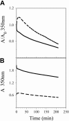
Fibrinolysis of α251 and control fibrin clots from decrease of turbidity. Clots were prepared and lysis was carried out as described in “Materials and methods.” Representative curves are shown. Solid line, control fibrin; broken line, α251 fibrin. (A) Change of turbidity as measured. (B) Change of turbidity normalized with respect to the initial turbidity of the clot.

References

    1. Mosesson MW, Hainfeld J, Wall J, Haschemeyer RH. Identification and mass analysis of human fibrinogen molecules and their domains by scanning transmission electron microscopy. J Mol Biol. 1981;153: 695-718. - PubMed
    1. Medved LV, Gorkun OV, Privalov PL. Structural organization of C-terminal parts of fibrinogen A alpha-chains. FEBS Lett. 1983;160: 291-295. - PubMed
    1. Weisel JW, Stauffacher CV, Bullitt E, Cohen C. A model for fibrinogen: domains and sequence. Science. 1985;230: 1388-1391. - PubMed
    1. Weisel JW, Medved L. The structure and function of the alpha C domains of fibrinogen. Ann N Y Acad Sci. 2001;936: 312-327. - PubMed
    1. Veklich YI, Gorkun OV, Medved LV, Nieuwenhuizen W, Weisel JW. Carboxyl-terminal portions of the alpha chains of fibrinogen and fibrin: localization by electron microscopy and the effects of isolated alpha C fragments on polymerization. J Biol Chem. 1993;268: 13577-13585. - PubMed

Publication types

MeSH terms