. 2005 Aug;26(7):1612-22.

Evaluation of the pathologic characteristics of excitotoxic spinal cord injury with MR imaging

Affiliations

Evaluation of the pathologic characteristics of excitotoxic spinal cord injury with MR imaging

Sara A Berens et al. AJNR Am J Neuroradiol. 2005 Aug.

Abstract

Background and purpose: Although high-resolution MR imaging is a valuable diagnostic tool, in vivo MR imaging has not yet been compared with in vitro MR imaging and histologic techniques following experimental spinal cord injury (SCI). The goal of the present study was to evaluate the feasibility of using in vivo MR imaging, in vitro MR imaging, and histologic techniques to study pathologic changes associated with excitotoxic SCI at a single time point. These results are important for future research using in vivo MR imaging to study the temporal profile of pathologic changes following SCI.

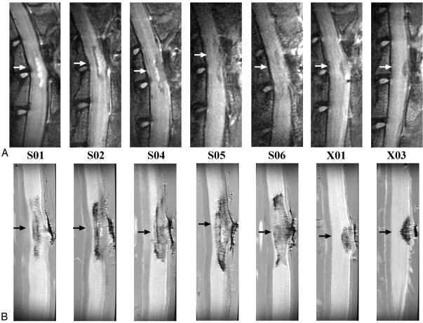
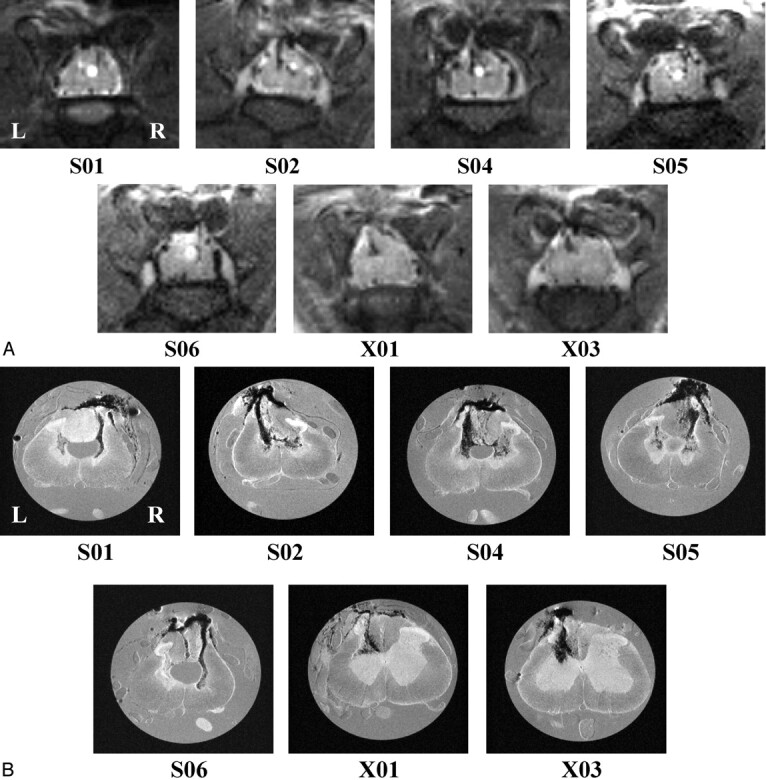
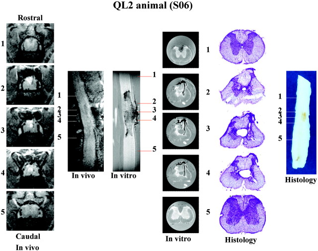
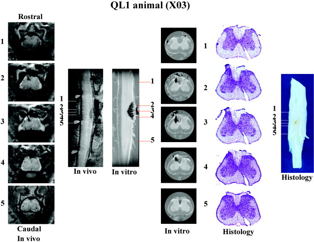
Methods: Rats received intraspinal injections of quisqualic acid at the T12-L2 spinal level. In vivo T1- and T2-weighted and dynamic contrast-enhanced MR images were collected 17-24 days postinjury. Once completed, spinal cords were removed and in vitro MR microscopy and histologic assessment were performed. MR images were collected using 4.7-T (in vivo) and 14.1-T magnets (in vitro).

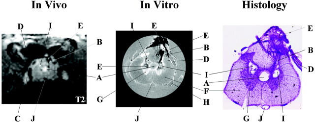
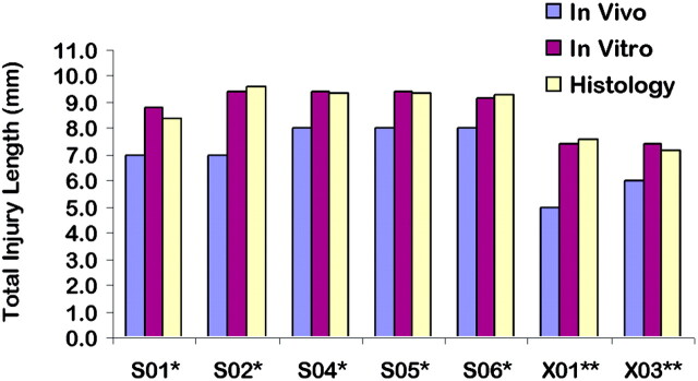
Results: Pathologic changes--including hemorrhage, neuronal loss, cavities, and central canal expansion--were visible in T2-weighted in vivo MR images. Evaluation of the blood-spinal cord barrier after injury with contrast agent enhancement showed no disruption at the time points evaluated. In vitro MR images and histologic evaluation confirmed pathologic details observed in vivo.

Conclusion: Results show that high-resolution in vivo MR imaging has the potential to be used in studying the progression of pathologic changes at multiple time points following SCI. This strategy may provide a way of studying structure-function relationships between therapeutic interventions and different pathologic characteristics of the injured spinal cord.


Figures

F<sc>ig</sc> 1.
Fig 1.
Representative pathologic findings in the same animal (S05) at the same location observed in vivo (T2-weighted, TR/effective − TE = 2000 ms/62.5 ms; NA = 6), in vitro (TR/TE = 2500 ms/20 ms; NA = 8), and in histologic sections. Many of the same pathologic details are visible with all three methods albeit with different levels of resolution. In vivo and in vitro images are displayed with image size = 0.92 cm × 0.77 cm (in vivo) and 0.50 cm × 0.50 cm (in vitro). Letters label the most distinctive anatomic and pathologic characteristics observed in the three images as follows: A indicates expanded central canal; B, injection track; C, dorsal root ganglion; D, dorsal root; E, hemorrhage at injury site; F, white matter; G, gray matter; H, ventral root; I, cavity; J, anterior spinal vessel. In vivo images show cavitation as hyperintense signals due to T2 weighting, whereas, on in vitro images, cavities appear isointense in these proton attenuation-weighted images.
F<sc>ig</sc> 2.
Fig 2.
A, In vivo (T2-weighted, TR/effective TE = 2000 ms/62.5 ms; NA = 6); and, B, in vitro (TR/TE = 2500 ms/20 ms; NA = 8) sagittal images for all QUIS-injured animals. Images are oriented with rostral at the top and dorsal on the right. Arrows represent the location of the epicenter of the injury. The epicenter was defined as the region of maximal pathologic damage. Displayed image size = 0.77 cm × 2.15 cm (in vivo) and 0.40 cm × 1.50 cm (in vitro). Animals designated S01, S02, S04, S05, and S06 received QL2, whereas animals designated X01 and X03 received QL1. The vertebral column and laminectomy site were easily observed in both in vivo and in vitro sagittal images.
F<sc>ig</sc> 3.
Fig 3.
A, In vivo (T2-weighted, TR/effective − TE = 2000 ms/62.5 ms; NA = 6); and, B, in vitro (TR/TE = 2500 ms/20 ms; NA = 8) transverse images for all QUIS-injured animals. Images are sampled from the epicenter of the injury (arrows in Fig 2) and show comparable pathologic changes between methods. See Fig 1 for labeling of anatomic and pathologic findings. Images are oriented with dorsal at the top. L, left side of cord; R, right side of cord, for all images. Displayed image size = 0.92 cm × 0.77 cm (in vivo) and 0.50 cm × 0.50 cm (in vitro). A, Hypointense signals correlated with hemorrhage (see “Histologic Findings”) and hyperintense signals correlated with the presence of fluid filled cavities. For example, notice the hyperintense expanded central canal in the cord of animals S01, S02, and S04–S06 and the hyperintense cavity in the gray matter on the right side of the cord in animal S02. B, Hypointense regions were observed bilaterally in the gray matter in all QL2 animals (S01, S02, and S04–S06), whereas, in QL1 animals, evidence of hemorrhage was observed only ipsilateral to the side of QUIS injection (X01 and X03). In addition, hypointense areas were observed in the dorsal columns in all the QUIS-injured animals except S01. Central canal expansion was clearly defined in four of the five QL2 animals (S01 and S04–S06), and injection tracks were visible in all injured animals. Cavity formation was also seen in all QL2 animals bilaterally.
F<sc>ig</sc> 4.
Fig 4.
Total injury lengths determined by using in vivo, in vitro, and histologic methods for each QUIS-injured animal. Total injury lengths in millimeters reflect the presence of excitotoxic tissue damage. No significant differences are observed between in vitro and histology (P > .05). Significant differences are observed between in vivo and in vitro/histology (P < .05), but these differences are within the range of section thickness difference between in vivo and in vitro images. A single asterisk denotes the QL2 animal, and double asterisks denote the QL1 animal.
F<sc>ig</sc> 5.
Fig 5.
Summary of in vivo, in vitro, and histologic data collected for a representative QL2 animal (S06). Representative sections (1–5) are sampled from the same rostrocaudal location in the spinal cord with each method used. Image sizes are the same as in Figs 2 and 3. See Fig 1, for labeling of anatomic and pathologic findings. Although sagittal images show the rostral-caudal extent of the injury, transverse sections provide greater detail of the pathologic changes associated with this injury model.
F<sc>ig</sc> 6.
Fig 6.
Summary of in vivo, in vitro, and histologic data collected for a representative QL1 animal (X03). Representative sections (1–5) are sampled with each method from the same rostrocaudal location in the spinal cord. Image sizes are the same as in Figs 2 and 3. See Fig 1, for labeling of anatomic and pathologic findings. Although sagittal images show the rostral-caudal extent of the injury, transverse sections provide greater detail of the pathologic changes associated with this injury model.

Comment in

References

    1. Yezierski RP. Pathophysiology and animal models of spinal cord injury pain. In: Yezierski RP, Burchiel K, eds. Spinal cord injury pain: assessment, mechanisms, management. Seattle: IASP Press;2002. :117–136
    1. Yezierski RP, Santana M, Park SH, Madsen PW. Neuronal degeneration and spinal cavitation following intraspinal injections of quisqualic acid. J Neurotrauma 1993;10:445–456 - PubMed
    1. Yezierski RP, Liu S, Ruenes GL, et al. Excitotoxic spinal cord injury: behavioral and morphological characteristics of a central pain model. Pain 1998;75:141–155 - PubMed
    1. Falconer JC, Narayana PA, Bhattacharjee MB, Liu SJ. Quantitative MRI of spinal cord injury in a rat model. Magn Reson Med 1994;32:484–491 - PubMed
    1. Ohta K, Fujimura Y, Watanabe M, Yato Y. Experimental study on MRI evaluation of the course of cervical spinal cord injury. Spinal Cord 1999;37:580–584 - PubMed

Publication types

MeSH terms

Substances