

Vacuum clefts of the vertebral bodies
- PMID: 16091506
- PMCID: PMC7975148
Vacuum clefts of the vertebral bodies
Abstract
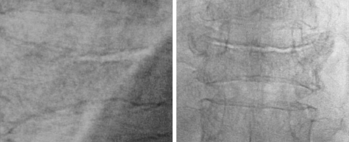
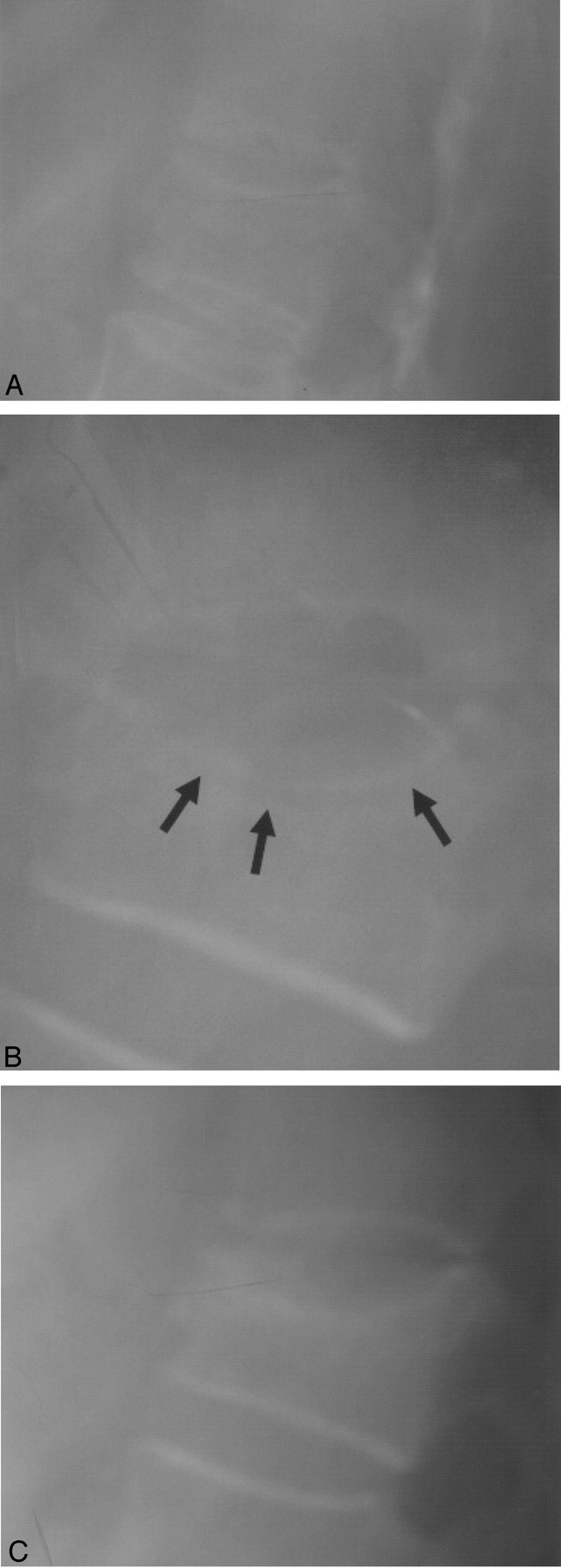
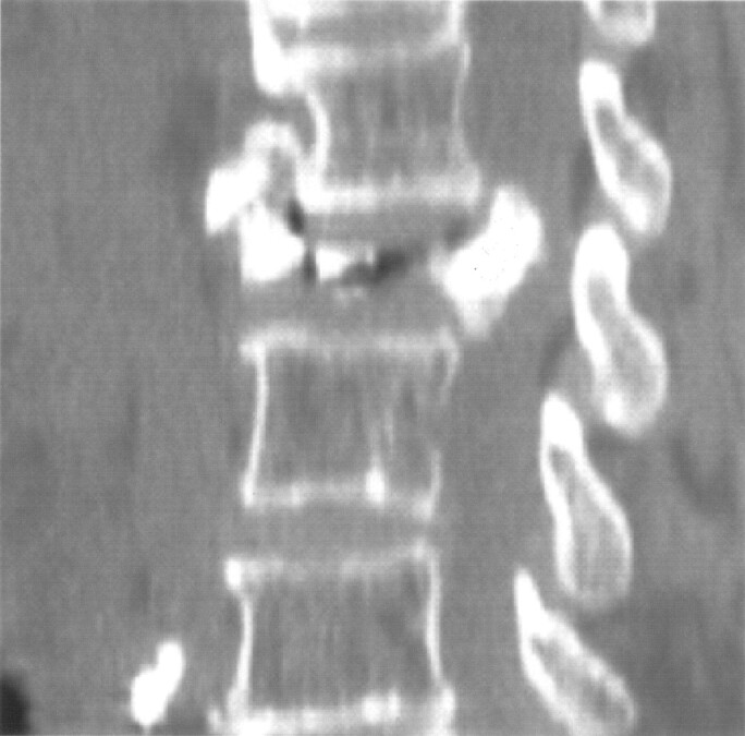
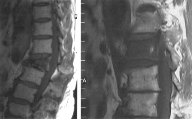
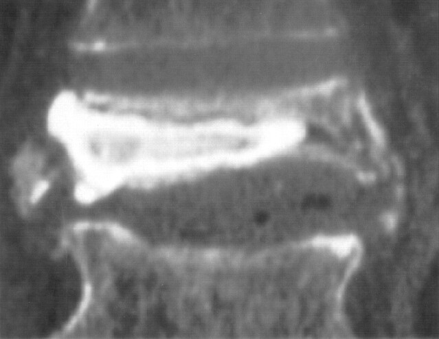
Background and purpose: The appearance of vacuum clefts (VCs) of the vertebral bodies has frequently been considered pathognomonic for avascular necrosis. Until recently, this was considered to be a rare finding that might indicate excessive motion at the fracture site. Our aim in this retrospective study was to determine the occurrence and location of these clefts in patients with osteoporotic vertebral fractures and evaluate the risk factors involved for developing these clefts in such patients.
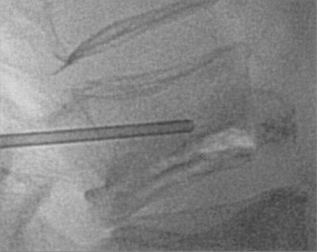
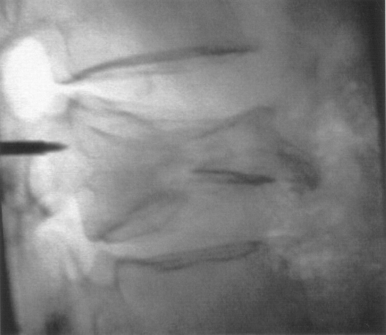
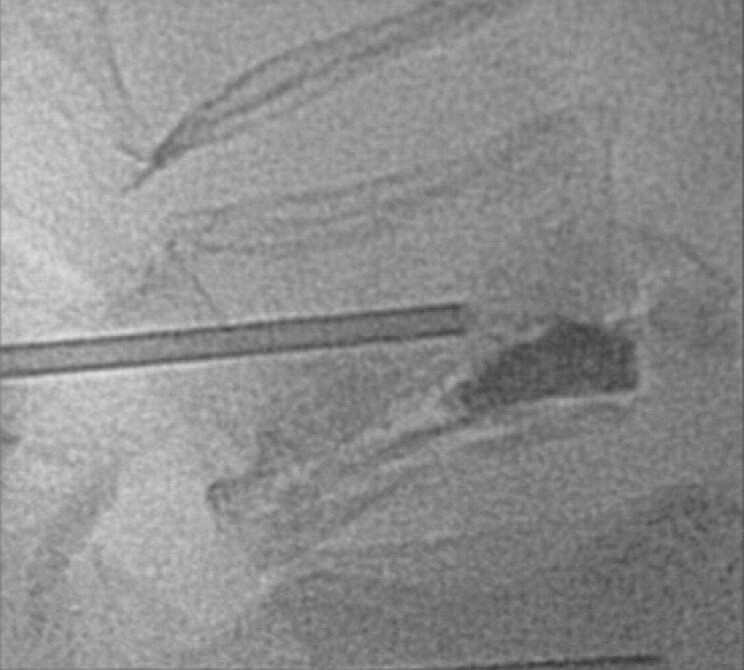
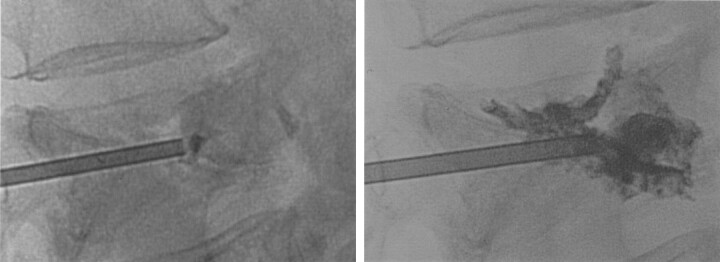
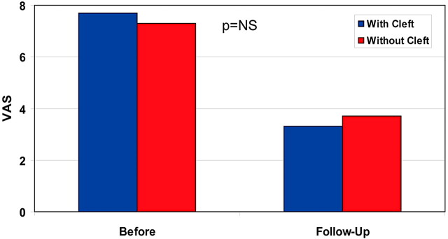
Methods: The records of 66 patients with 101 painful osteoporotic vertebral fractures who were treated by vertebroplasty in our department were reviewed. All the fractures with VCs were collected. Age, sex, degree of deformity, and extent of degenerative changes in the adjacent disk space were compared with those found in the patients without clefts.
Results: VCs were found in 26 fractured vertebrae of 26 patients. They were significantly more common in elderly men who had deformed fractures located at the thoracolumbar junction, when compared with fractures without clefts, especially when degenerative changes were observed in the adjacent disk space.
Conclusion: This study suggests that VCs, which have long been considered pathognomonic for avascular necrosis (Kümmell disease), are not rare and most probably represent fracture nonunion. Elderly patients who have deformed fractures at the thoracolumbar area have a higher risk for developing clefts, mainly when there is degeneration of the adjacent disk space.
Figures
References
-
- Maldague BE, Noel HM, Malghem JJ. The intravertebral vacuum cleft: a sign of ischemic vertebral collapse. Radiology 1978;29:23–29 - PubMed
-
- Theodorou DJ. The intravertebral vacuum cleft sign. Radiology 2001;221:787–788 - PubMed
-
- Kumpan W, Salomonowitz E, Seidl G, Wittich GR. The intravertebral vacuum phenomenon. Skeletal Radiol 1986;15:444–447 - PubMed
-
- McKiernan F, Faciszewski T. Intravertebral clefts in osteoporotic vertebral compression fractures. Arthritis Rheum 2003;48:1414–1419 - PubMed
-
- McKiernan F, Jensen R, Faciszewski T. The dynamic mobility of vertebral compression fractures. J Bone Miner Res 2003;18:24–29 - PubMed
MeSH terms
LinkOut - more resources
Full Text Sources
Medical