. 2005 Aug;26(7):1646-54.

Spinal cord schistosomiasis: MR imaging appearance with surgical and pathologic correlation

Affiliations

Spinal cord schistosomiasis: MR imaging appearance with surgical and pathologic correlation

Sahar Saleem et al. AJNR Am J Neuroradiol. 2005 Aug.

Abstract

Background and purpose: Spinal cord involvement is a rare manifestation of schistosomiasis. We describe the MR imaging findings of spinal cord schistosomiasis in correlation with surgery and pathology.

Methods: We report eight cases of spinal cord schistosomiasis. All patients were men (mean age, 16.7 years) with neurologic manifestations who had been referred for spinal MR imaging. In all cases, spinal masses were surgically removed. MR imaging findings were correlated with surgery and pathology.

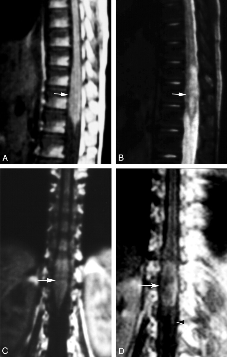
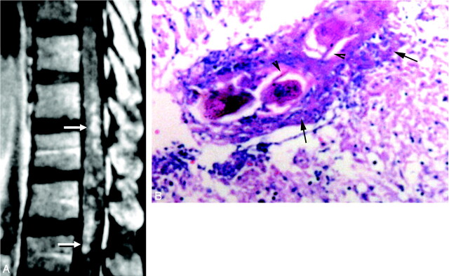
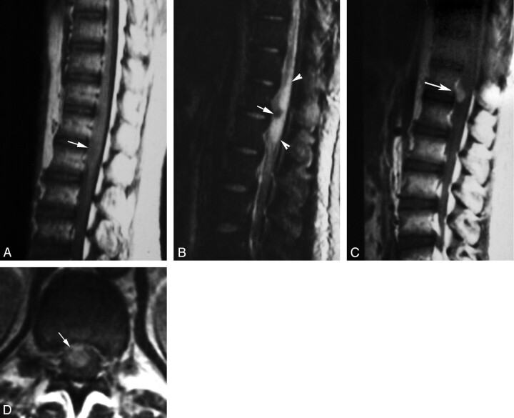
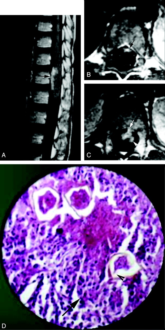
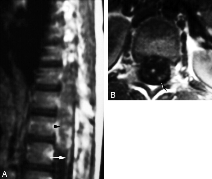
Results: MR imaging showed moderate expansion of the distal spinal cord in all cases. Abnormalities were isointense to cord in T1 and patchy hyperintense in T2-weighted spin-echo images (n = 8). Three forms of contrast enhancement were recognized: (1) intramedullary nodular (n = 8); (2) peripheral (n = 8); and (3) linear radicular (n = 4). Total gross surgical removal of masses by using the Cavitron ultrasonic surgical aspirator was possible in six cases. Diagnosis was established by identification of ova in histopathologic studies: Schistosoma mansoni (n = 3), S. hematobium (n = 1), and uncertain species (n = 4). Intramedullary nodular enhancement was correlated to multiple schistosomiasis microtubercles. Peripheral enhancing lesions correlated to thickened leptomeninges infested by chronic granulomatous inflammatory cells and schistosoma eggs. Linear radicular enhancement correlated with thickened resected nervous roots infested by granulomatous cells and schistosoma eggs.

Conclusion: Multinodular intramedullary contrast enhancement of the distal cord enabled correct presumptive preoperative MR imaging diagnosis of spinal schistosomiasis in three cases. Accurate diagnosis, through recognition of its MR imaging appearance, allows early treatment and better prognosis of spinal cord schistosomiasis.


Figures

F<sc>ig</sc> 1.
Fig 1.
Localization of spinal cord schistosomiasis. MR imaging of dorso-lumbar spine of case 3. A, Sagittal T1-weighted SE (TR/TE 530/20 ms) image shows moderate expansion of the distal cord and conus medullaris (arrow). The lesion is isointense to the cord. B, Sagittal T2-weighted SE (TR/TE 5800/95 ms) image. The lesion has heterogeneous hyperintense signal intensity (arrow). C, Coronal T1-weighted SE MR images (TR/TE 520/20 ms) shows the expanded distal cord and conus medullaris (arrow). D, Postcontrast coronal image, in which the lesion is well delineated by contrast enhancement (arrow). Note the associated linear enhancement of cauda equina nerve roots (arrowhead).
F<sc>ig</sc> 2.
Fig 2.
Diffuse nodular enhancement form in spinal cord schistosomiasis. A, Postcontrast sagittal T1-weighted SE MR image (TR/TE 520/20 ms) of case 5, showing multiple small intramedullary enhancing nodules diffusely involving the distal thoracic cord and conus medullaris (arrows). B, High-power photomicrograph stained with H & E (×250) of the patient, showing multiple granulomas. Schistosoma ova are seen in the midst of the granulomas surrounded by chronic inflammatory cells (arrows). Arrowheads point to the lateral spines, characteristic of S mansoni ova. The surrounding neural tissues are edematous and infiltrated by chronic inflammatory cells.
F<sc>ig</sc> 3.
Fig 3.
Masslike nodular enhancement form in spinal cord schistosomiasis. MR imaging of dorso-lumbar spine of case 6. A, Sagittal T1-weighted SE (TR/TE: 530/20 ms) image showing mild expansion of the distal cord and conus medullaris by a poorly defined iso- to hypointense lesion (arrow). B, Sagittal T2-weighted SE (TR/TE 5800/95 ms) image showing a hypointense lesion (arrow) surrounded by extensive perilesional hyperintense edema (arrowheads). C, Postcontrast sagittal T1-weighted SE MR images (TR/TE: 520/20 ms). The lesion shows a solitary enhancing nodule (arrow). D, Postcontrast axial image (TR/TE: 520/20 ms) documenting the intramedullary location of the enhancing spinal lesion (arrow).
F<sc>ig</sc> 4.
Fig 4.
Peripheral enhancement form of spinal cord schistosomiasis. MR imaging of dorso-lumbar spine and histopathologic slide of case 2. A, Postcontrast sagittal image (TR/TE 520/20 ms) shows peripheral and intramedullary enhancing lesions on the anterior surface of the distal spinal cord (arrow). B, Postcontrast axial image (TR/TE 520/20 ms), clearly showing the peripheral (meningeal) enhancement on the anterior surface of the cord (arrow). C, Postcontrast axial image, which is inferior to that shown in B, shows a small peripheral enhancing lesion on the anterior cord surface (arrow) in association with an underlying intramedullary cord enhancement (arrowhead). D, High-power photomicrograph stained with H & E (×250) showing infiltration of the resected neural tissues by chronic inflammatory cells (arrow) surrounding multiple schistosoma ova. One of the ova shows the terminal spine characteristic of S hematobium (arrowhead).
F<sc>ig</sc> 5.
Fig 5.
Radicular enhancement form in spinal cord schistosomiasis. Postcontrast T1-weighted images (TR/TE 520/20 ms) of case 1. A, Sagittal image shows linear enhancement of cauda equina nerve roots (arrow). Note the associated diffuse nodular intramedullary and peripheral enhancement of the distal cord and conus medullaris (arrowhead). B, Axial image documenting the thickened enhancing cauda equina nerves (arrow).

Similar articles

Cited by

References

    1. World Health Organization. Public health impact of schistosomiasis. Bull WHO 1993;71:623–657 - PMC - PubMed
    1. Mahmoud AA. Trematodes (schistosomiasis, other flukes). In: Mandell GL, Douglas RG, Bennet JE, eds. Principles and practice of infectious diseases. 2nd ed. New York: Wiley Medical;1985. :1573–1579
    1. El Banhawy A, Elwan O, Taher Y. Bilharzial granuloma of the conus medullaris and cuada equina. Paraplegia 1972;10:172–180 - PubMed
    1. Luyendijk W, Lindeman J. Schistosomiasis (Bilharziasis) mansoni of the spinal cord simulating an intramedullary tumor. Surg Neurol 1975;4:457–460 - PubMed
    1. Dar J, Zimmerman RR. Schistosomiasis of the spinal cord. Surg Neurol 1977;8:416–418 - PubMed

MeSH terms