

Case Reports
Double-balloon remodeling of wide-necked aneurysms distal to the circle of Willis
Affiliations
- PMID: 16091528
- PMCID: PMC7975178
Item in Clipboard
Case Reports
Double-balloon remodeling of wide-necked aneurysms distal to the circle of Willis
AJNR Am J Neuroradiol.
2005 Aug.
Abstract
We describe a remodeling technique in which two compliant balloon catheters were used to treat large, wide-necked bifurcation aneurysms distal to the circle of Willis in two patients with subarachnoid hemorrhage. Although vasospasm and interaction between the constrained catheters in the small distal arteries added to the complexity of the procedure, placement of a balloon catheter in each side branch prevented coil encroachment and enabled embolization of these aneurysms.
Figures
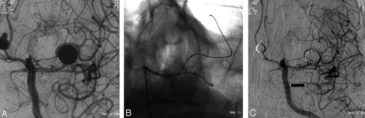
Case 1. A, Left internal carotid arteriogram (working projection) before bypass of the aneurysm shows that both of the trunks of middle cerebral artery arise from the aneurysm. Microcatheter in the middle cerebral artery and in the anterior communicating artery aneurysm are noted. B, Digital radiograph shows both catheters (HyperForm; Micro Therapeutics, Inc) in the superior and inferior trunks. Microcatheter tip is in the aneurysm. C, Left internal carotid arteriogram after embolization of the anterior communicating artery aneurysm shows satisfactory obliteration of both aneurysms.
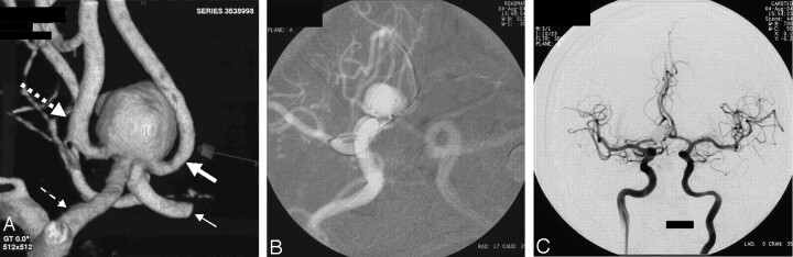
Case 2. A, Aneurysm viewed from its posterior aspect on a 3D angiogram, which shows involvement of the origins of both A2 segments. Small dashed arrow indicates left A1; large dashed arrow, left A2; small solid arrow, right A1; and large solid arrow, right A2. B, Live road-map image shows the crossed pattern of catheters extending from A1 segments to the contralateral A2 segments and the catheter tip in the aneurysm. C, Bilateral internal carotid arteriograms at the end of the procedure demonstrate a small neck remnant.
Similar articles
-
Embolization of a wide-necked basilar bifurcation aneurysm by double-balloon remodeling using HyperForm compliant balloon catheters.J Clin Neurosci. 2009 Apr;16(4):560-2. doi: 10.1016/j.jocn.2008.05.031. Epub 2009 Feb 23. J Clin Neurosci. 2009. PMID: 19231201
-
Balloon-assisted coil placement in wide-neck bifurcation aneurysms by use of a new, compliant balloon microcatheter.AJNR Am J Neuroradiol. 2003 Jun-Jul;24(6):1222-5. AJNR Am J Neuroradiol. 2003. PMID: 12812958 Free PMC article.
-
Ballonn-assisted Guglielmi detachable coiling of wide-necked aneurysma: Part II--clinical results.Neurosurgery. 1999 Sep;45(3):531-7; discussion 537-8. doi: 10.1097/00006123-199909000-00024. Neurosurgery. 1999. PMID: 10493375
-
Modified balloon assisted coil embolization for the treatment of intracranial and cervical arterial aneurysms using coaxial dual lumen balloon microcatheters: initial experience.J Neurointerv Surg. 2014 Nov;6(9):704-7. doi: 10.1136/neurintsurg-2013-010936. Epub 2013 Oct 23. J Neurointerv Surg. 2014. PMID: 24153339
-
Complex intracranial aneurysms: combined operative and endovascular approaches.Neurosurgery. 1998 Dec;43(6):1304-12; discussion 1312-3. doi: 10.1097/00006123-199812000-00020. Neurosurgery. 1998. PMID: 9848843 Review.
Cited by
-
Endovascular treatment of wide-neck middle cerebral artery aneurysms with stents: a review of 16 cases.AJNR Am J Neuroradiol. 2010 May;31(5):940-6. doi: 10.3174/ajnr.A1931. Epub 2009 Dec 31. AJNR Am J Neuroradiol. 2010. PMID: 20044506 Free PMC article.
-
Evaluation of a neck-bridge device to assist endovascular treatment of wide-neck aneurysms of the anterior circulation.AJNR Am J Neuroradiol. 2008 Jan;29(1):73-8. doi: 10.3174/ajnr.A0767. Epub 2007 Oct 10. AJNR Am J Neuroradiol. 2008. PMID: 17928379 Free PMC article. Clinical Trial.
-
Remodeling Technique in the Treatment of Intracranial Aneurysms: Indications, Limits and Non-indications.Interv Neuroradiol. 2008 Sep 1;14 Suppl 1(Suppl 1):52-9. doi: 10.1177/15910199080140S110. Epub 2008 Oct 9. Interv Neuroradiol. 2008. PMID: 20557775 Free PMC article. No abstract available.
-
Comaneci plus Balloon-assisted Embolization of Ruptured Wide-necked Cerebral Aneurysms.Clin Neuroradiol. 2022 Sep;32(3):773-782. doi: 10.1007/s00062-021-01115-0. Epub 2022 Jan 18. Clin Neuroradiol. 2022. PMID: 35041011
-
Wide neck bifurcation aneurysms: what is the optimal endovascular treatment?J Neurointerv Surg. 2021 May;13(5):e9. doi: 10.1136/neurintsurg-2021-017459. Epub 2021 Mar 15. J Neurointerv Surg. 2021. PMID: 33722965 Free PMC article. No abstract available.
References
-
- Henkes H, Kirsch M, Mariushi W, Miloslavski E, Brew S, Kuhne D. Coil treatment of a fusiform upper basilar trunk aneurysm with a combination of “kissing” neuroform stents, TriSpan-, 3D- and fibered coils, and permanent implantation of the microguidewires. Neuroradiology 2004;46:464–468 - PubMed
-
- Moret J, Pierot L, Boulin A, Castaings L. “Remodelling” of the arterial wall of the parent vessel in the endovascular treatment of intracranial aneurysms. Neuroradiology 1994;36(suppl 1):83
-
- Takahashi A. Neck plastic intra-aneurysmal GDC embolisation with double protective balloons: method of multiple guiding catheter introduction. Intervent Neuroradiol 1998;4:177–179 - PubMed
Publication types
MeSH terms
LinkOut - more resources
Full Text Sources
Other Literature Sources
Medical