Angiographic assessment of pial collaterals as a prognostic indicator following intra-arterial thrombolysis for acute ischemic stroke
- PMID: 16091531
- PMCID: PMC7975179
Angiographic assessment of pial collaterals as a prognostic indicator following intra-arterial thrombolysis for acute ischemic stroke
Abstract
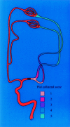
Background and purpose: This study examines whether anatomic extent of pial collateral formation documented on angiography during acute thromboembolic stroke predicts clinical outcome and infarct volume following intra-arterial thrombolysis, compared with other predictive factors.
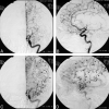
Methods: Angiograms, CT scans, and clinical information were retrospectively reviewed in 65 consecutive patients who underwent thrombolysis for acute ischemic stroke. Clinical data included age, sex, time to treatment, National Institutes of Health Stroke Scale (NIHSS) score on presentation of symptoms, NIHSS score at the time of hospital discharge, and modified Rankin scale score at time of hospital discharge. Site of occlusion, scoring of anatomic extent of pial collaterals before thrombolysis, and recanalization (complete, partial, or no recanalization) were determined on angiography. Infarct volume was measured on CT scans performed 24-48 hours after treatment.
Results: Fifty-three patients (82%) qualified for review. Both infarct volume and discharge modified Rankin scale scores were significantly lower for patients with better pial collateral scores than those with worse pial collateral scores, regardless of whether they had complete (P < .0001) or partial (P = .0095) recanalization. Adjusting for other factors, regression analysis models indicate that the infarct volume was significantly larger (P < .0001) and modified discharge Rankin scale score and discharge NIHSS score significantly higher for patients with worse pial collateral scores. Similarly, adjusting for other factors, the infarct volume was significantly lower (P = .0006) for patients with complete recanalization than patients with partial or no recanalization.
Conclusions: Evaluation of pial collateral formation before thrombolytic treatment can predict infarct volume and clinical outcome for patients with acute stroke undergoing thrombolysis independent of other predictive factors. Thrombolytic treatment appears to have a greater clinical impact in those patients with better pial collateral formation.
Figures


References
-
- Zeumer H, Freitag HJ, Zanella F, et al. Local intra-arterial fibrinolytic therapy in patients with stroke: urokinase versus recombinant tissue plasminogen activator (r-TPA). Neuroradiology 1993;35:159–162 - PubMed
-
- National Institute of Neurological Disorders and Stroke rt-PA Stroke Study Group. Tissue plasminogen activator for acute ischemic stroke. N Engl J Med 1995;333:1581–1587 - PubMed
-
- Theron J, Coskun O, Huet H, et al. Local intraarterial thrombolysis in the carotid territory. Intervent Neuroradiol 1996;2:111–126 - PubMed
-
- Furlan A, Higashida R, Wechsler L, et al. Intra-arterial prourokinase for acute ischemic stroke: the PROACT II study: a randomized controlled trial. JAMA 1999;282:2003–2011 - PubMed
-
- Viroslav AB, Hoffman JC. The use of computed tomography in the diagnosis of stroke. Heart Dis Stroke 1993;2:299–307 - PubMed
MeSH terms
LinkOut - more resources
Full Text Sources
Medical
