. 2005 Dec 8;24(55):8167-75.
doi: 10.1038/sj.onc.1208950.

The nuclear receptor liver receptor homolog-1 is an estrogen receptor target gene

Affiliations

The nuclear receptor liver receptor homolog-1 is an estrogen receptor target gene

Jean-Sébastien Annicotte et al. Oncogene. .

Abstract

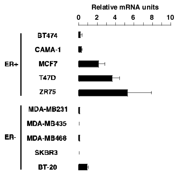
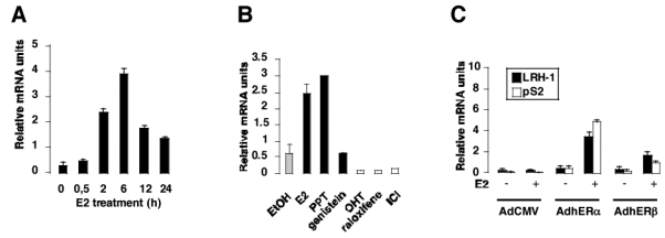
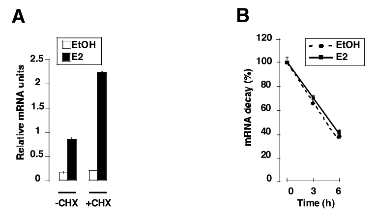
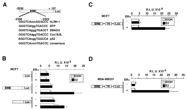
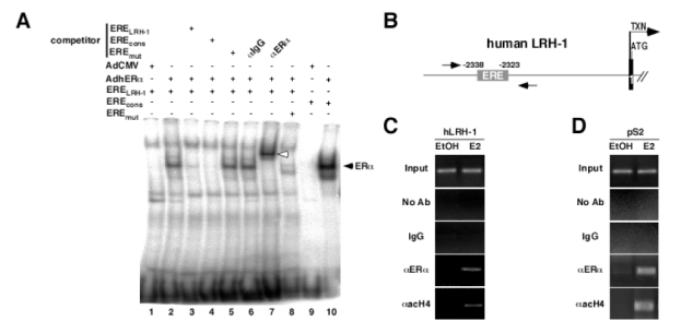
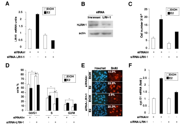
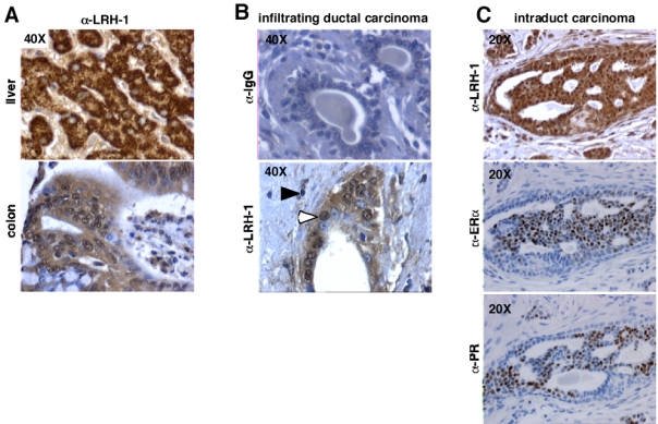
Liver receptor homolog-1 (LRH-1) is a nuclear receptor previously known to have distinct functions during mouse development and essential roles in cholesterol homeostasis. Recently, a new role for LRH-1 has been discovered in tumor progression, giving LRH-1 potential transforming functions. In order to identify critical factors stimulating LRH-1 expression leading to deregulated cellular proliferation, we studied its expression and its regulation in several breast cancer cell lines. We observed that LRH-1 expression was increased in estrogen receptor (ER) alpha expressing cell lines, whereas weak-to-no expression was found in nonexpressing ERalpha cell lines. In MCF7, LRH-1 expression was highly induced after treatment with 17beta-estradiol (E2). This transcriptional regulation was the result of a direct binding of the ER to the LRH-1 promoter, as demonstrated by gelshift and chromatin immunoprecipitation assays. Interestingly, siRNA-mediated inactivation of LRH-1 decreased the E2-dependent proliferation of MCF7 cells. Finally, LRH-1 protein expression was detected by immunohistochemistry in tumor cells of human mammary ductal carcinomas. Altogether, these data demonstrate that LRH-1 is transcriptionally regulated by the ER alpha and reinforce the hypothesis that LRH-1 could exert potential oncogenic effects during breast cancer formation.


Figures

Figure 1
Figure 1. LRH-1 is specifically expressed in ER+ breast cancer cell lines
Total mRNA was isolated from several ERα expressing (ER+) and non expressing (ER−) breast cancer cell lines cultured in phenol red free medium containing 10% DCC-FCS. RNA was reverse transcribed as described in the Experimental Procedures. Levels of LRH-1 mRNA were determined by Q-PCR and normalized to RS9.
Figure 2
Figure 2. E2 regulates LRH-1 expression in MCF7 cells
A- MCF7 cells were stripped from endogenous steroids and cultured in phenol red free medium containing 3% DCC-FCS, with E2 at a concentration of 10−8M. Cells were harvested at different time and processed for mRNA extraction. LRH-1 expression was measured by Q-PCR, and data are expressed relative to RS9. B- Cells were stripped as described in A and incubated with the control vehicle (EtOH), ERα agonists (E2, PPT), ER partial agonist (genistein) or ER antagonists (OHT, raloxifene, ICI) at 10−8M. After 24 hours, RNA was extracted and LRH-1 expression was measured as described in A. C- The ER- cell line MDA-MB231 was stripped from endogenous steroids as described and infected with non-recombinant adenovirus (AdCMV) or with adenovirus containing the human ERα (AdERα) or β (AdERβ) cDNA. After infection, cells were treated with the vehicle (−) or E2 10−8M (+) for 48 hours. Cells were then harvested and processed for RNA extraction and Q-PCR as described. LRH-1 and pS2 expressions were measured as described in A.
Figure 3
Figure 3. E2 regulation of LRH-1 is not secondary to an intermediate protein or higher mRNA stability
A- MCF7 were treated (+CHX) or not (−CHX) for 1 hour with cycloheximide (25μ/ml) before adding the vehicle (EtOH) or E2 (10−8M). Cells were scrapped 15 hours after E2 treatment and RNA was extracted as described in the Experimental Procedures section. Quantification of LRH-1 expression was performed by Q-PCR and standardized to RS9 levels. The results are means of three independent experiments. B- MCF7 cells were treated with ethanol (EtOH) or E2 (10−8M) for 24 hours and then incubated for different times (0, 3, 6 hours) with actinomycin D (3μg/ml) before RNA extraction. LRH-1 expression was measured by Q-PCR, normalized to RS9 and the results are expressed as 100% of LRH-1/RS9 RNA levels without actinomycin D treatment (0 hour). The results are means of three independent experiments.
Figure 4
Figure 4. ERα regulates LRH-1 promoter activity
A- Computational analysis of a 2.5 kb fragment of the regulatory region of the human LRH-1 gene demonstrating the presence of a potential binding site for ER homologous to the ERE present in the EFP, ABAG9, Cox7A2L, pS2 genes, except for one base pair. A consensus sequence for the ERE is also represented. B- MCF7 cells were transfected with (+) or without (−) an expression vector encoding ERα and the pGL3 basic vector, the hLRH-1 reporter vector or a deletion mutant devoid of the ERE. Cells were then treated with the control vehicle ethanol (EtOH) or E2 10−8M for 24 hours. After treatment, cells were lysed and assayed for luciferase activity. C- The ERELRH-1-Tk-Luc construct was transfected in MCF7 in the presence (+) or absence (−) of ERα. Cells were treated with ethanol (EtOH) or E2 at a concentration of 10−8M, harvested after 24 hours and processed for luciferase activity as described in B. D- The same construct as in C was cotransfected in the ER- cell line MDA-MB231 with an empty vector or a vector encoding ERα. Cells were stimulated and assayed for luciferase activity as in B.
Figure 5
Figure 5. ERα binds to the ERE present in the hLRH-1 promoter
A- EMSA showing binding of ERα to the ERE present in the hLRH-1 promoter. Whole cell nuclear extracts from MDA-MB231 infected with a non recombinant adenovirus (lane 1 and 9) or an adenovirus encoding the human ERα (lane 2 to 8, 10) were incubated with radiolabeled ERELRH-1 (lane 1 to 7), EREmut (lane 8) or EREcons (positive control, lane 9 and 10), in the presence or absence of wild-type (competitor ERELRH-1, lane 3), consensus (competitor EREcons, lane 4) or mutated (competitor EREmut, lane 5) cold probe. Incubation of an anti-ERα antibody resulted in a supershifted band (lane 7, white arrow), whereas no modification in ERα binding was observed with IgG (lane 6). B- Schematic representation of the 5′ region of the human LRH-1 gene. The ERELRH-1 is indicated. Transcription (arrow, TXN) and translation (ATG) initiation sites are shown. Amplimers are highlighted by the arrows. C–D ChIP assay demonstrating binding of ERα to the human LRH-1 and pS2 promoters. Cross-linked chromatin from MCF7 treated with the vehicle (EtOH) or E2 10−8M (E2) for 48 hours were incubated without antibody (No Ab), IgG or antibodies against human ERα (aERa) and acetylated histone H4 (aacH4). Immunoprecipitates were analyzed by PCR and positive controls (Input) are shown.
Figure 6
Figure 6. siRNA-mediated inhibition of LRH-1 expression decreases E2 effect on cell proliferation
A- RNA levels of LRH-1 are decreased in MCF7 expressing a siRNA-LRH-1 in the absence or presence of E2. Q-PCR results were normalized to RS9. B- Protein levels of LRH-1 are decreased in MCF7 expressing a siRNA-LRH-1. An anti-β-actin antibody was used as a loading control. C- Cell proliferation was quantified as described in the Experimental Procedures section. Proliferation of MCF7 cells expressing a LRH-1 siRNA is decreased compared to MCF7 expressing an irrelevant siRNA in the absence (EtOH) or presence of estradiol (E2). D- FACS analysis of propidium iodide-stained cells comparing the cell cycle profile of MCF7 cells stably expressing an irrelevant or LRH-1 siRNA, in the absence (EtOH) or presence of estradiol (E2). E- BrdU incorporation in MCF7 siRNAirr and siRNA-LRH-1 cells (Texas red fluorescence, right) compared to the total number of nuclei (Hoechst staining, left). The average percentage of BrdU-labeled cells form five independent field is indicated. F- siRNA-mediated inhibition of LRH-1 decreases cyclin D1 mRNA expression in MCF7 cells. RNA levels of the Gl cyclin were determined by Q-PCR and normalized to RS9.
Figure 7
Figure 7. LRH-1 is expressed in tumor cells of mammary infiltrating ductal carcinomas
A- We used 5 μm formalin-fixed paraffin-embedded liver and colon tissue sections, as a positive control for immunostaining. A strong immunoreactivity was detected on these tissues. B- As negative controls, mouse IgGs were used instead of the primary antibody on breast tissue section. No staining was observed in these conditions. C- LRH-1 immunostaining (open arrowheads) is detected in tumor cells of mammary ductal carcinomas, with a nuclear and cytoplasmatic localization. Inflammatory stromal regions surrounding carcinomas contain some lymphocytes positively stained for LRH-1 (filled arrowheads).

References

    1. Ali S, Coombes RC. Nat Rev Cancer. 2002;2:101–12. - PubMed
    1. Annicotte JS, Fayard E, Swift GH, Selander L, Edlund H, Tanaka T, Kodama T, Schoonjans K, Auwerx J. Mol Cell Biol. 2003;23:6713–24. - PMC - PubMed
    1. Bardin A, Boulle N, Lazennec G, Vignon F, Pujol P. Endocr Relat Cancer. 2004;11:537–51. - PMC - PubMed
    1. Botrugno OA, Fayard E, Annicotte JS, Haby C, Brennan T, Wendling O, Tanaka T, Kodama T, Thomas W, Auwerx J, Schoonjans K. Mol Cell. 2004;15:499–509. - PubMed
    1. Castoria G, Migliaccio A, Bilancio A, Di Domenico M, de Falco A, Lombardi M, Fiorentino R, Varricchio L, Barone MV, Auricchio F. Embo J. 2001;20:6050–9. - PMC - PubMed

Publication types