

Construction of tissue microarrays from prostate needle biopsy specimens
- PMID: 16091762
- PMCID: PMC2361582
- DOI: 10.1038/sj.bjc.6602726
Construction of tissue microarrays from prostate needle biopsy specimens
Abstract
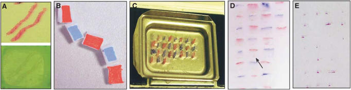
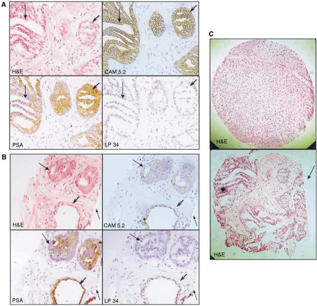
Needle biopsies are taken as standard diagnostic specimens for many cancers, but no technique exists for the high-throughput analysis of multiple individual immunohistochemical (IHC) markers using these samples. Here we present a simple and highly reliable technique for constructing tissue microarrays (TMAs) from prostatic needle biopsies. Serial sectioning of the TMAs, called 'Checkerboard TMAs', facilitated expression analysis of multiple proteins using IHC markers. In total, 100% of the analysed biopsies within the TMA both preserved their antigenicity and maintained their morphology. Checkerboard TMAs will allow the use of needle biopsies (i) alongside other tissue specimens (trans-urethral resection of prostates and prostatectomies in the case of prostate cancer) in clinical correlation studies when searching for new prognostic markers, and (ii) in a diagnostic context for assessing expression of multiple proteins in cancers from patients prior to treatment.
Figures
References
-
- Bastacky S, Cieply K, Sherer C, Dhir R, Epstein JI (2004) Use of interphase fluorescence in situ hybridisation in prostate needle biopsy specimens with isolated high-grade prostatic intraepithelial neoplasia as a predictor of prostate adenocarcinoma on follow-up biopsy. Hum Pathol 35: 281–289 - PubMed
-
- Battifora H, Mehta P (1990) The checkerboard tissue block. An improved multitissue control block. Lab Invest 63: 722–724 - PubMed
-
- Bostwick DG (1997) Evaluating prostate needle biopsy: therapeutic and prognostic importance. CA Cancer J Clin 47: 297–319 - PubMed
-
- Bubendorf L, Kononen J, Koivisto P, Schraml P, Moch H, Gasser TC, Willi N, Mihatsch MJ, Sauter G, Kallioniemi OP (1999) Survey of gene amplifications during prostate cancer progression by high-throughout fluorescence in situ hybridisation on tissue microarrays. Cancer Res 59: 803–806 - PubMed
-
- Cormier JN, Pollock RE (2004) Soft tissue sarcomas. CA Cancer J Clin 54: 94–109 - PubMed
Publication types
MeSH terms
LinkOut - more resources
Full Text Sources
Other Literature Sources
Medical
