. 1992 Jun 19;256(5064):1684-7.
doi: 10.1126/science.256.5064.1684.

Hydrogen exchange measurement of the free energy of structural and allosteric change in hemoglobin

Affiliations

Hydrogen exchange measurement of the free energy of structural and allosteric change in hemoglobin

S W Englander et al. Science. .

Abstract

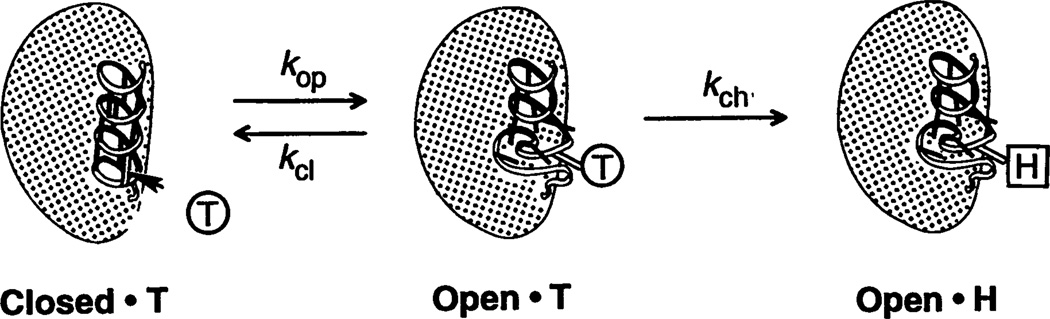
The inability to localize and measure the free energy of protein structure and structure change severely limits protein structure-function investigations. The local unfolding model for protein hydrogen exchange quantitatively related the free energy of local structural stability with the hydrogen exchange rate of concerted sets of structurally related protons. In tests with a number of modified hemoglobin forms, the loss in structural free energy obtained locally from hydrogen exchange results matches the loss in allosteric free energy measured globally by oxygen-binding and subunit dissociation experiments.


Figures

Fig. 1
Fig. 1
Diagrammatic representation of a local unfolding reaction. A section of helix is pictured to unfold transiently and reversibly, breaking a set of contiguous hydrogen bonds. Protein NH (pictured with tritium label) can exchange with solvent hydrogens only from the open state, the occupation of which is determined by the unfolding (opening) equilibrium constant, Kop. The HX rate determined by this sequence can generally be represented as in Eq. 1. kex=kopkch[OH]/(kop+kcl+kch[OH])Kopkch[OH] Here kop and kcl are opening and closing rate constants and kCh[OH] is the chemical exchange rate characteristic of the freely exposed hydrogen (specific base–catalyzed above pH 3). In a real case, the NHs in an unfolding reaction may not all exchange at the same rate. Some dispersion in rates is imposed by chemical inductive effects of nearest neighbor side chains (15). Physical effects may also intervene, such as partial protection of some residues in the open state. Equation 2a (where R is the gas constant and T is temperature) recognizes that Kop is determined by the equilibrium free energy of the opening reaction (ΔG°op). ΔG°op=RTlnKop δΔG°op=RTδlnKop=RTln(kex,1/kex,2) A change in the free energy of unfolding (δΔG°OP) will produce a multiplicative change in Kop and therefore a multiplicative change in the HX rate of all the hydrogens in the set, as in Eq. 2b. Equation 2b assumes that the change in structure or interaction represented by δΔG°op affects the native state only and not the energy level of the transiently unfolded state.
Fig. 2
Fig. 2
Hydrogen-tritium exchange curves for allosterically sensitive peptide NH at (A) the α-chain NH2-terminus and (B) the β-chain COOH-terminus of human Hb (pH 7.4, 0°C, 0.1 M NaCI, 0.1 M phosphate, ±20 mM pyrophosphate, and 5 mM ferrous ammonium sulfate; Hb concentration ~0.5 mM in tetramers). HX data are shown for deoxy Hb and several modified forms. For each modified Hb, the HX time scale is shown multiplied by a factor that brings its exchange curve into consonance with the unmodified deoxy Hb curve (two-exponential fit). (A): (○), HbA, ×1; (▲), NES, ÷2; (●), oxy Hb, ×9. (B): (○), HbA, ×1; (■), no PPi, ×4; (▲), NES, ×8, (●), oxy Hb, ×750. The multiplying factor was computer-determined. In these experiments, allosterically sensitive sites were selectively tritium-labeled by the functional labeling method (–5). Hemoglobin was initially exchanged-in in the fast-exchanging, oxy form in tritiated water (THO) for 35 min. Both allosterically sensitive and insensitive NH sites that exchange in 35 min become labeled. Hemoglobin was then deoxygenated (sodium dithionite), and exchange-out was immediately initiated by removing free 3H1HO (short gel filtration passage through a 1 cm by 6 cm column of G25 fine Sephadex). At allosterically insensitive sites tritium label is soon lost, because exchange-out proceeds at the same rate as did exchange-in. At allosterically sensitive sites, now in their slow form, tritium is locked into a more slowly exchanging form. Thus, after a short exchange-out time the hemoglobin sample selectively retains label essentially at allosterically sensitive sites. Tritium label remaining unexchanged at various protein locations as a function of exchange-out time was measured by a fragment separation method (6). Hemoglobin samples prepared as just described were taken after various exchange-out times, placed into slow HX conditions (pH 3, 0°C) by passage through a 3-cm Sephadex column (<1 min), and proteolyzed with pepsin (5 min). The fragments were separated by high-pressure liquid chromatography (~20 min), and the label on each fragment was counted by liquid scintillation. The allosterically sensitive NHs at the α-chain NH2-terminus were measured on the fragment α1–29 (only four of the five sensitive NHs in the set are labeled in the 35-min exchange-in). The four sensitive NHs at the β-chain COOH-terminus (Fig. 3) were recovered and measured on the fragment β130–146. A correction was made for the calibrated loss of label during the fragment separation analysis (19 and 33%, respectively, for the α- and β-chain sets). A low-level background curve due to labeled allosterically insensitive sites was generated and subtracted as described (4).
Fig. 3
Fig. 3
Interactions in the vicinity of the allosterically sensitive peptide NHs at the β-chain COOH-terminus (7). The β-chain heme is shown coordinated to the proximal histidine (His-92) on the F-FG segment. This segment is joined to the COOH-terminal segment by a number of allosterically sensitive bonds, indicated by shading. Each modification tested breaks one or more of these cross-links, as indicated in Table 1. The hydrogen-bonded NHs believed to account for the HX behavior measured at the α-chain COOH-terminus (Fig. 2B) are indicated by dotted lines.

Similar articles

Cited by

References

    1. Berger A, Linderstrøm-Lang K. Arch. Biochem. Biophys. 1957;69:106. - PubMed
    2. Eigen M. Angew. Chem. Int. Ed. Engl. 1964;3:1.
    3. Hvidt A, Nielsen SO. Adv. Protein Chem. 1966;21:287. - PubMed
    1. Englander SW. Ann. N.Y Acad. Sci. 1975;244:10. - PubMed
    1. Englander JJ, Rogero JR, Englander SW. J. Mol. Biol. 1983;169:325. - PubMed
    1. Englander SW, Englander JJ. In: Structure and Dynamics of Nucleic Acids and Proteins. Clementi E, Sarma RH, editors. Guilderland, NY: Adenine; 1983. pp. 421–434.
    2. Rogero JR, Englander SW, Englander JJ. Methods Enzymol. 1986;131:L508. - PubMed
    3. Ray J, Englander SW. Biochemistry. 1986;25:3000. - PubMed
    4. Louie G, Tran T, Englander JJ, Englander SW. J. Mot. Biol. 1988;201:755. - PubMed
    5. Louie G, Englander JJ, Englander SW. ibid. :765. - PubMed
    6. McKinnie RE, Englander JJ, Englander SW. Chem. Phys. 1991;158:283.
    1. Functional labeling is now generically termed hydrogen exchange labeling in parallel with analogous methods being used to study kinetic refolding intermediates, equilibrium unfolding intermediates, and protein interaction sites. For a review, see Englander SW, Mayne L. Annu. Rev. Biophys. Biomol. Struct. 1992;21:243.

Publication types

LinkOut - more resources