Characterization of insulin-like-growth factor II (IGF II) mRNA positive hepatic altered foci and IGF II expression in hepatocellular carcinoma during diethylnitrosamine-induced hepatocarcinogenesis in rats
- PMID: 16092956
- PMCID: PMC1199609
- DOI: 10.1186/1477-3163-4-12
Characterization of insulin-like-growth factor II (IGF II) mRNA positive hepatic altered foci and IGF II expression in hepatocellular carcinoma during diethylnitrosamine-induced hepatocarcinogenesis in rats
Abstract
Background: Insulin-like-growth factor II (IGF II) has been implicated in the pathogenesis of neoplasm of different tissues, including liver of rats and men. This growth factor is believed to exert its effect during cellular proliferation. During the process of development of hepatocellular carcinoma (HCC), different hepatic altered foci appear. They are believed to be the putative precursors of HCC in rats and in men. Thus, to study the role of the gene in a defined model of hepatocarcinogenesis was the target to elucidate its role in various cancer phenotypes during the entire development stage of cancer, right from earlier preneoplastic lesions to HCC.
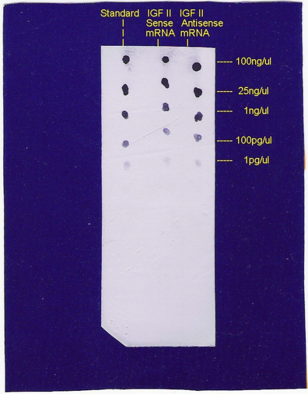
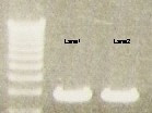
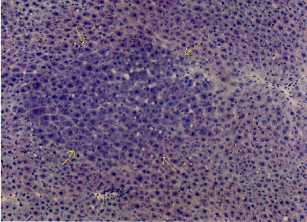
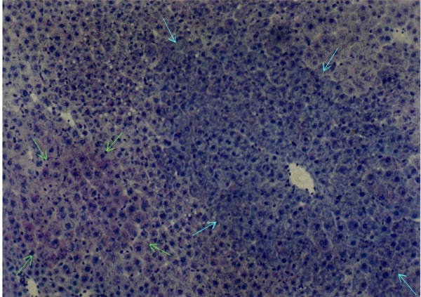
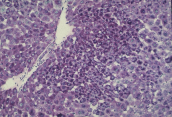
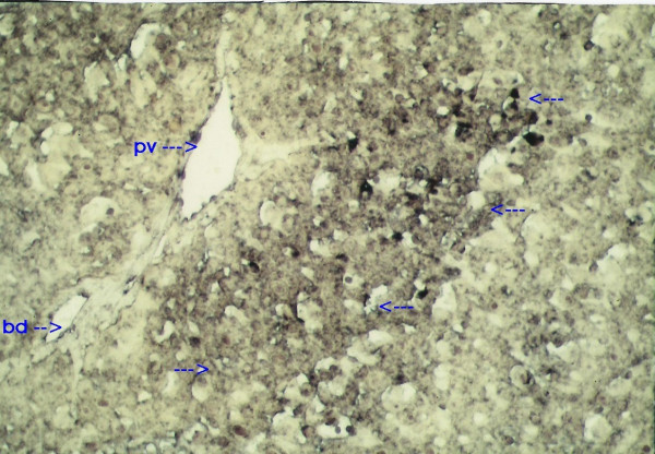
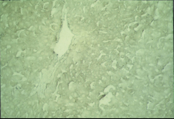
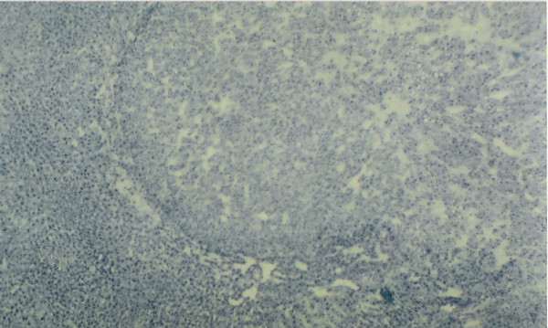
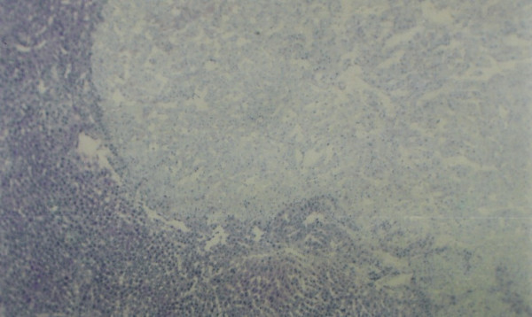
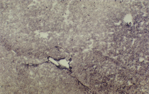
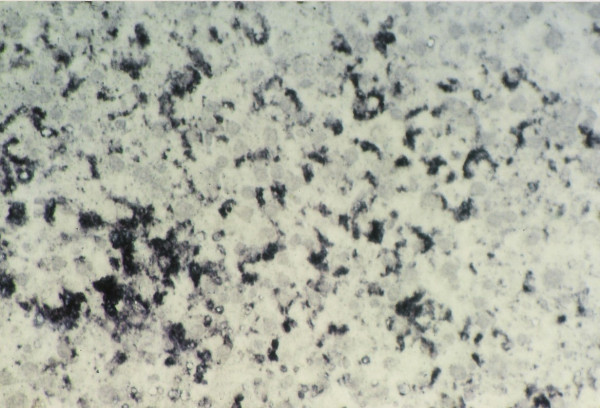
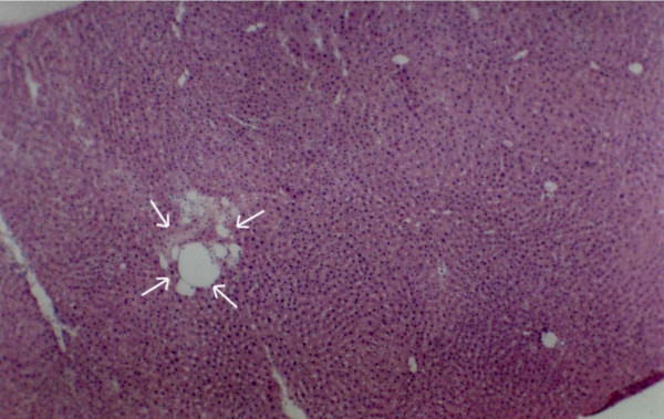
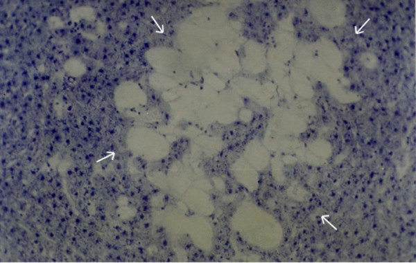
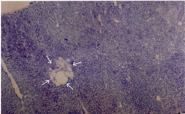
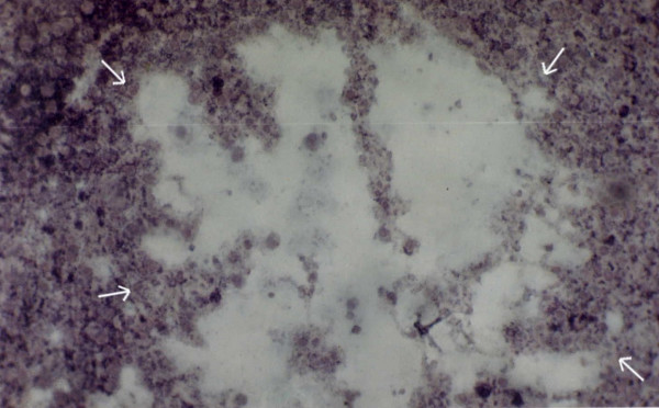
Methods: Antisense in situ hybridization technique was used here to characterize the type(s) of foci in which IGF II mRNA had expressed during the development of hepatocarcinogenesis-induced by diethylnitrosamine and promoted by phenobarbital in rats. Various focal lesions have been categorized depending on the stages and sizes along with IGF II expression patterns in them. Immunohistochemical detection for proliferating cell nuclear antigen (PCNA) was made to detect the role of the gene in preneoplastic and neoplastic cellular proliferation.
Results: IGF II expression was located in the glycogen-storage acidophilic cell foci maximally followed by mixed cell lesions and the least in basophilic lesions. The expression of IGF II was found to be predominant in the HCC. The expression of gene was also located at the peripheral cells of spongiosis hepatis which are believed to be the precursor of ito cell carcinoma. It was noted that there is a direct correlation between IGF II expression and immunohistochemical detection for PCNA.
Conclusion: It may be concluded that IGF II gene expression plays an important role during the development of neoplasia and the gene expresses in the sequence of events leading from glycogen-rich-acidophilic lesions to glycogen poor basophilic lesions to HCC with an expression pattern of "high-low-high" in terms of degree of expression. Moreover, the essential role of the gene at the immediate initiation stage of carcinogenesis (first few weeks) and during HCC development cannot be ignored. Thus this expression can be used as a suitable marker for very early detection of the cancerous process and can save numbers of future cancer victims by very early detection of this disease.
Figures
References
-
- Daughaday WH, Rotwein P. Insulin-like-growth factors I and II peptide, messenger ribonucleic acid and gene structures, serum and tissue concentrations. Endocrinol Rev. 1989;10:68–91. - PubMed
-
- Brown AL, Graham DE, Nissley SP, Hill DJ, Strain AJ, Rechler MM. Developmental regulation of insulin like growth factor II mRNA in different rat tissues. J Biol Chem. 1986;261:445–462. - PubMed
-
- Cariani E, Lasserre C, Seurin D, Hamelin B, Kemeny F, Franco D, Czech MP, Ullrich A, Brechot C. Differential expression of insulin like growth factor II mRNA in human primary liver cancer, benign tumors and liver cirrhosis. Cancer Res. 1998;48:6844–6849. - PubMed
LinkOut - more resources
Full Text Sources
Miscellaneous