. 2005 Oct 21;280(42):35458-68.
doi: 10.1074/jbc.M506648200. Epub 2005 Aug 10.

Suppression of death receptor-mediated apoptosis by 1,25-dihydroxyvitamin D3 revealed by microarray analysis

Affiliations

Suppression of death receptor-mediated apoptosis by 1,25-dihydroxyvitamin D3 revealed by microarray analysis

Xiaohui Zhang et al. J Biol Chem. .

Abstract

Recent studies suggest that growth inhibition by 1,25-dihydroxyvitamin D3 represents an innovative approach to ovarian cancer therapy. To understand the molecular mechanism of 1,25-dihydroxyvitamin D3 action, we profiled the hormone-induced changes in the transcriptome of ovarian cancer cells using microarray technology. More than 200 genes were identified to be regulated by 1,25-dihydroxyvitamin D3. Reverse transcription-PCR analyses confirmed the regulation of a group of apoptosis-related genes, including the up-regulation of the decoy receptor that inhibits tumor necrosis factor-related apoptosis-inducing ligand (TRAIL) action, TRAIL receptor 4, and the down-regulation of Fas, the receptor that mediates the action of Fas ligand. The regulation was further confirmed at the protein level. Consistent with the regulation of the death receptors, pretreatment with 1,25-dihydroxyvitamin D3 decreased apoptosis induced by TRAIL and Fas ligand. Because persistent 1,25-dihydroxyvitamin D3 treatment has been shown to induce apoptosis in ovarian cancer, the hormone appears to exert a dual effect on the death of ovarian cancer cells. Knockdown of TRAIL receptor 4 by RNA interference or ectopic expression of Fas relieved the suppressive effect of 1,25-dihydroxyvitamin D3, showing that molecular manipulation of death receptors is a viable approach to overcome the protective effect of 1,25-dihydroxyvitamin D3 on the apoptosis of ovarian cancer. These strategies may allow ovarian cancer patients to benefit from therapy with both 1,25-dihydroxyvitamin D3 and ligands for death receptors, such as TRAIL, shown to selectively induce apoptosis in cancer but not normal cells.


Figures
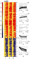
FIGURE 1
FIGURE 1. Gene expression profiles in OVCAR3 cells after 1,25-(OH)2D3 treatment
RNA isolated from OVCAR3 cells treated with 1,25-(OH)2D3 for 0, 8, 24, or 72 h was subjected to microarray analyses. Three independent analyses (I–III) were performed in Affymetrix U133A chips. A total of 96 genes with less than 5% false detective rate in SAM analysis (delta = 0.653) and at least 2.5-fold changes were selected for clustering analysis (GeneSpring software, Silicon Genetics). Genes are presented in rows. The time of 1,25-(OH)2D3 treatment and the number of array analyses are indicated above and below the column. Up- and down-regulated genes are shown with a colorimetric scale to indicate the relative expression levels. Numbers 1–5 show five different clusters identified by gene tree clustering.
FIGURE 2
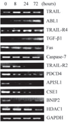
FIGURE 2. Verification of the regulation by 1,25-(OH)2D3 at mRNA level of apoptosis-related genes identified in microarray analyses
OVCAR3 cells were treated for the indicated times, and RNA was prepared and subjected to RT-PCR analyses with primers specific for different genes. Glyceraldehyde-3-phosphate dehydrogenase (GAPDH) control was included in each PCR analysis and representative data are shown. PDCD4, programmed cell death 4; CSE1, cellular apoptosis susceptibility 1; BNIP2, BCL2 adenovirus E1B 19-kDa interacting protein 2; HDAC1, histone deacetylase 1; AP5IL1, API5-like 1; TGF-β, transforming growth factor-β.
FIGURE 3
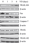
FIGURE 3. Regulation at the protein level of apoptotic genes along the death receptor pathway by 1,25-(OH)2D3
OVCAR3 cells were treated with 10−7 m 1,25-(OH)2D3 (VD) for the indicated times and the levels of TRAIL-R4, Fas, caspase-7, TRAIL, and TRAIL-R2 protein expression were determined by immunoblotting. β-Actin blot was included to show even loading.
FIGURE 4
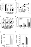
FIGURE 4. Suppression of TRAIL-induced apoptosis by 1,25-(OH)2D3 in OVCAR3 cells
A, cells were pretreated with 1,25-(OH)2D3 (VD) or vehicle (EtOH) for 3 days. Equal numbers of treated cells were plated into 96-well plates and further treated with the indicated concentrations of TRAIL for 24 h. MTT assays were performed and absorption at 595 nm (A595 nm) was determined. Eight samples were analyzed in parallel for each data point and the experiments were reproduced twice. Percentages of viable cells were calculated relative to the A595 values of cells before TRAIL treatment (pretreated cells), which was set as 100%. *, p < 0.01 when compared with the corresponding values from cells pretreated with vehicle. B, cells were pretreated similarly as in A and subsequently treated with or without 100 ng/ml TRAIL for the indicated times before MTT assays were performed. Percentages of viable cells were calculated by dividing A595 values with the corresponding value before treatment for each group. *, p < 0.01 when compared with the group treated with TRAIL following vehicle. C and D, cells were pretreated similarly as in A followed by secondary treatment with 100 ng/ml TRAIL for the indicated times. Apoptosis of treated cells was determined by flow cytometry after Annexin V staining. A representative profile of the flow cytometry analysis is shown in C. The apoptotic index from two independent analyses is shown in D. E, effect of 1,25-(OH)2D3 on TRAIL-induced caspase-7 activation. Cells were treated with 10−7 m 1,25-(OH)2D3 or EtOH for 0, 1, 3, or 6 days followed by treatment with 100 ng/ml TRAIL for 4 h. Capase-7 activity was determined as described under “Materials and Methods.” F, effect of 1,25-(OH)2D3 on TRAIL-induced caspase-3 activation. Cells were treated as in E and caspase-3 activity was measured. FITC, fluorescein isothiocyanate.
FIGURE 5
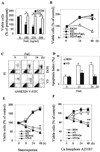
FIGURE 5. Selective suppression of Fas ligand (FasL)-induced apoptosis by 1,25-(OH)2D3 in OVCAR3 cells
A–D, cells were treated and MTT (panels A and B) and apoptotic (panels C and D) assays were performed as described in the legend to Fig. 4, panels A–D. The difference is that the cells were treated with FasL instead of TRAIL. For panel A, cells were treated for 48 h and, for panels B–D, cells were treated with 250 ng/ml FasL. A and B, *, p < 0.01 when compared with the corresponding values obtained from cells pretreated with vehicle. E and F, cells pretreated with 10−7 m 1,25-(OH)2D3 or vehicle for 3 days were treated with 1 µm staurosporin or 0.5 µm calcium ionophore A23187 for the indicated times. MTT assays were performed, and percentages of viable cells were calculated by dividing A595 values with the corresponding value before treatment. *, p < 0.01 when compared with cells treated with vehicle followed by staurosporin. **, p < 0.05 when compared with cells treated with vehicle followed by A23187. FITC, fluorescein isothiocyanate.
FIGURE 6
FIGURE 6. Suppression of TRAIL-induced apoptosis by 1,25-(OH)2D3 in multiple cancer cell lines
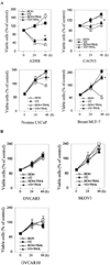
A and B, cells pretreated similarly as in Fig. 4A were subsequently treated with or without 100 ng/ml TRAIL for the indicated times before MTT assays were performed with the exception that LNCaP cells were treated with 800 ng/ml TRAIL. Percentages of viable cells were calculated by dividing A595 values with the corresponding value before treatment. *, p < 0.01 when compared with cells treated with TRAIL following vehicle.
FIGURE 7
FIGURE 7. Suppression of the death receptor-mediated apoptosis by 1,25-(OH)2D3 is eliminated by molecular manipulation of TRAIL-R4 and Fas
OVCAR3 cells pretreated with 10−7 m 1,25-(OH)2D3 or vehicle were transfected with TRAIL-R4-specific siRNA (A and B) or pCMV-Fas together with pEGFP plasmids (C and D). Six h post-transfection, cells were placed in fresh medium containing 1,25-(OH)2D3 or vehicle. 48 h later, cells were harvested and immunoblotting analysis was performed to determine TRAIL-R4 or Fas protein levels (panels A and C). For apoptosis analyses, cells were treated with 100 ng/ml TRAIL for 4 h (panel B) or 250 ng/ml Fas ligand for 12 h before fixation (panel D). TRAIL-induced apoptosis was determined by flow cytometry after Annexin-V staining (B). Fas ligand-induced apoptosis was scored by counting the percentage of GFP-positive cells undergoing nuclear condensation (D).

Similar articles

Cited by

References

    1. Webb AR, Kline L, Holick MF. J. Clin. Endocrinol. Metab. 1988;67:373–378. - PubMed
    1. Muir C, Waterhouse J, Mack T, Powell J, Whelan S. Cancer Incidence in Five Continents, International Agency for Research on Cancer Control (IARC), Scientific Publication No. 88. Vol. V. Lyon, France: IARC; 1987.
    1. Devesa SS, Grauman MA, Blot WJ, Pennello GA, Hoover RN, Fraumeni JF., Jr . Atlas of Cancer Mortality in the United States. Bethesda, MD: National Institutes of Health Publication No. 99-4564, NCI, National Institutes of Health; 1999.
    1. Jiang F, Li P, Fornace AJ, Jr, Nicosia SV, Bai W. J. Biol. Chem. 2003;278:48030–48040. - PubMed
    1. Miettinen S, Ahonen MH, Lou YR, Manninen T, Tuohimaa P, Syvala H, Ylikomi T. Int. J. Cancer. 2004;108:367–373. - PubMed

Publication types

MeSH terms