. 2005 Sep;96(4):625-38.
doi: 10.1093/aob/mci215. Epub 2005 Aug 10.

Rice: sulfide-induced barriers to root radial oxygen loss, Fe2+ and water uptake, and lateral root emergence

Affiliations

Rice: sulfide-induced barriers to root radial oxygen loss, Fe2+ and water uptake, and lateral root emergence

Jean Armstrong et al. Ann Bot. 2005 Sep.

Abstract

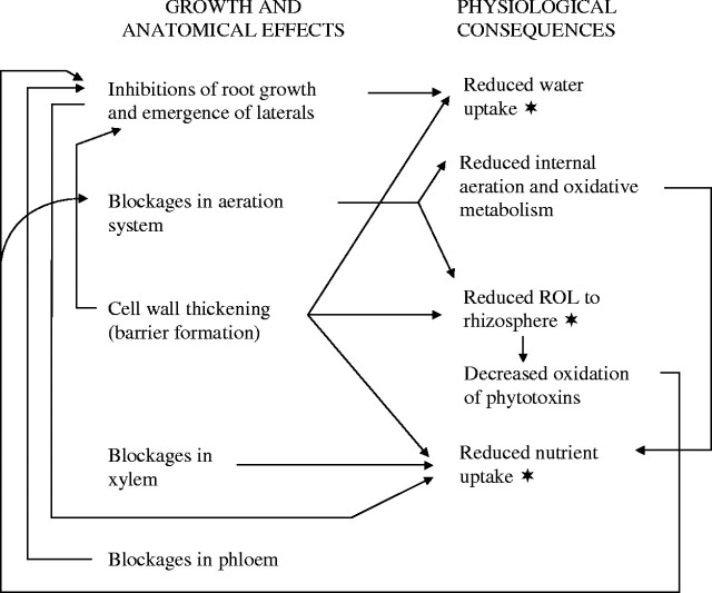
Background and aims: Akagare and Akiochi are diseases of rice associated with sulfide toxicity. This study investigates the possibility that rice reacts to sulfide by producing impermeable barriers in roots.

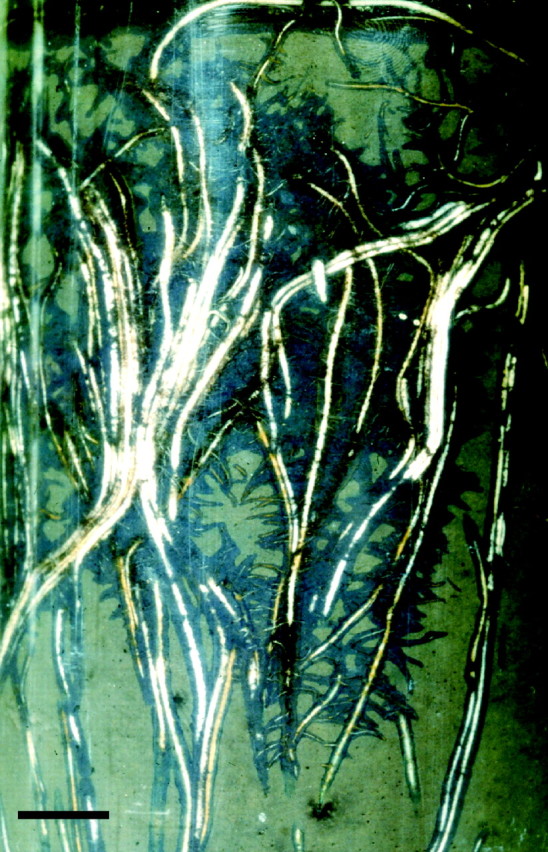
Methods: Root systems of rice, Oryza sativa cv. Norin 36, were subjected to short-term exposure to 0.174 mm sulfide (5.6 ppm) in stagnant solution. Root growth was monitored; root permeability was investigated in terms of polarographic determinations of oxygen efflux from fine laterals and the apices of adventitious roots, water uptake, anatomy and permeability to Fe2+ using potassium ferricyanide.

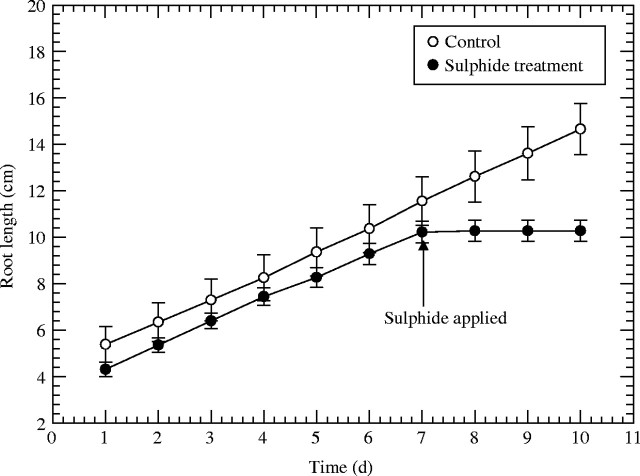
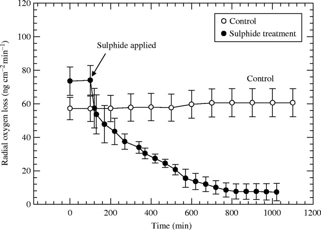
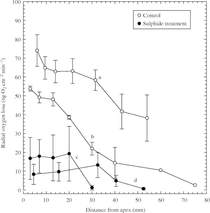
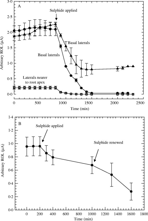
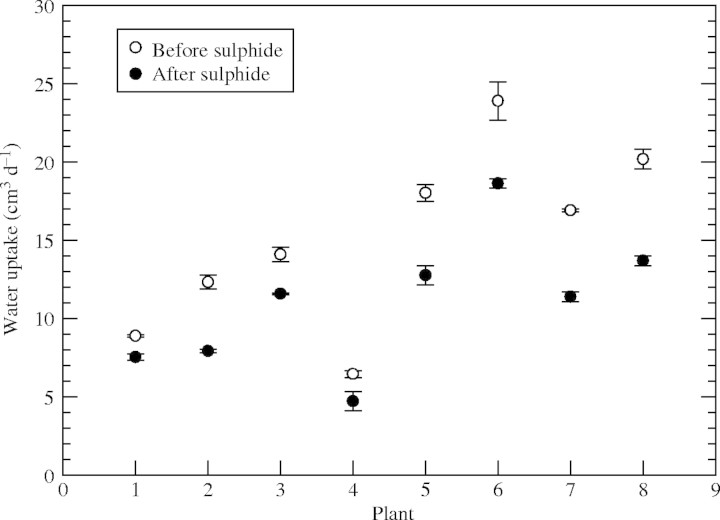
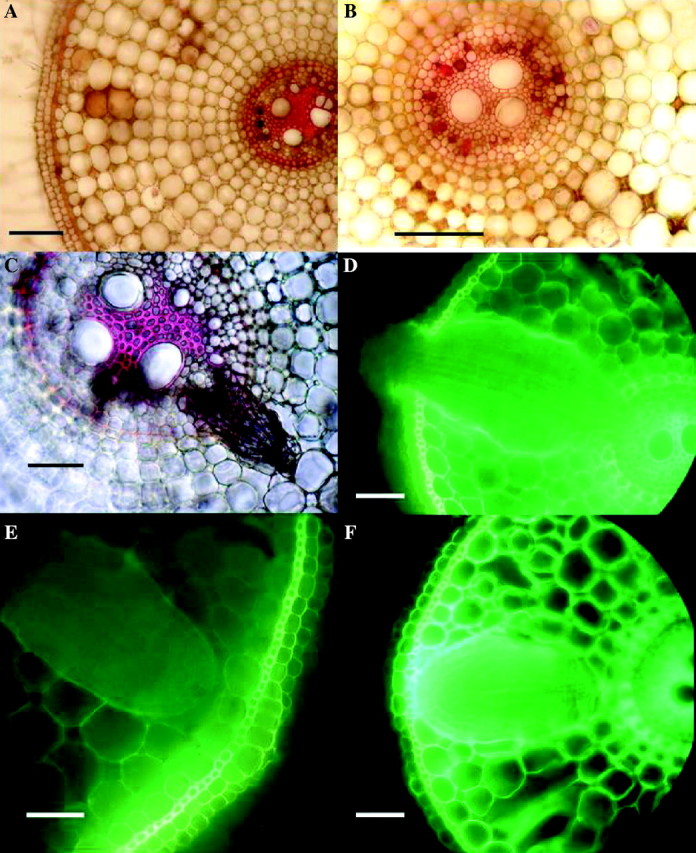
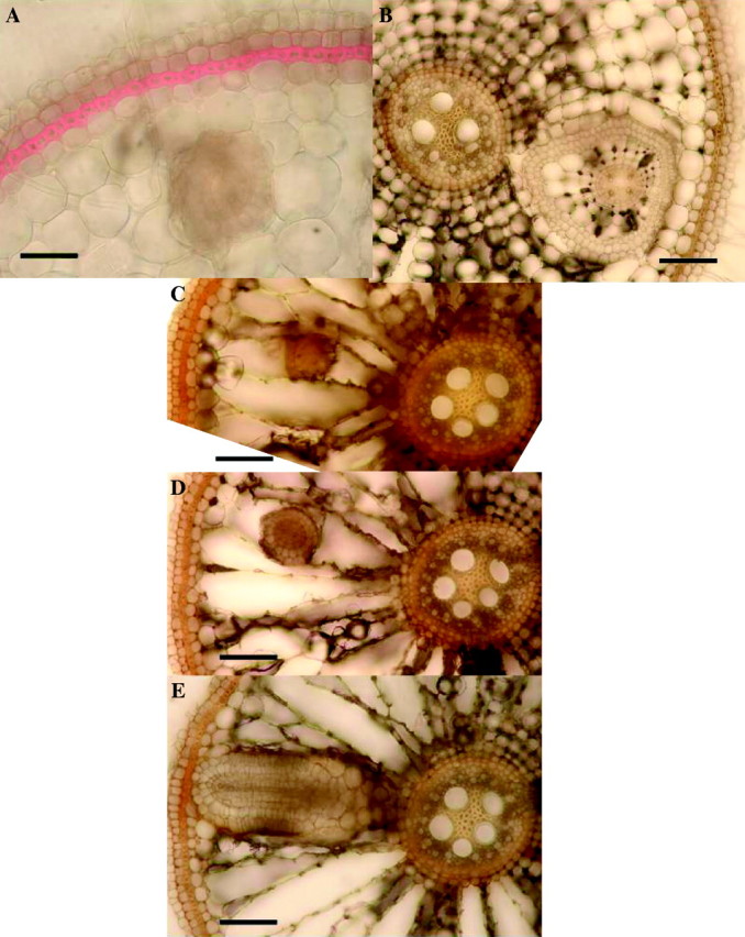
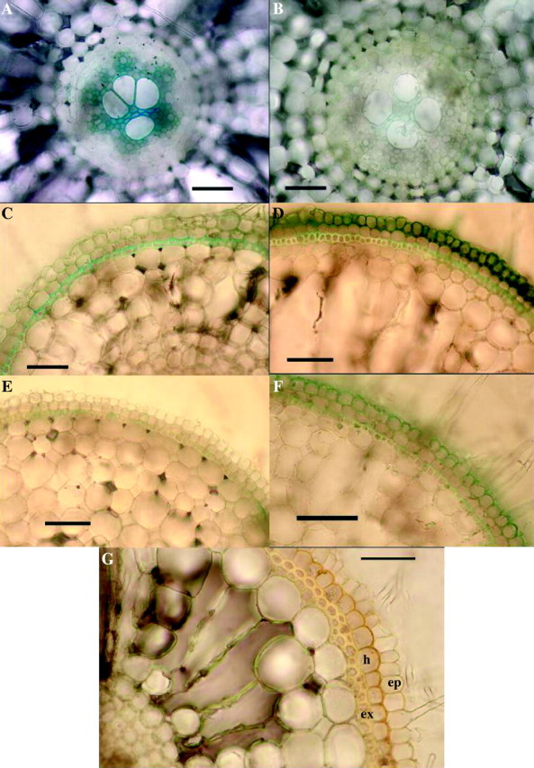
Key results: Both types of root responded rapidly to the sulfide with immediate cessation of growth, decreased radial oxygen loss (ROL) to the rhizospheres and reduced water uptake. Profiles of ROL measured from apex to basal regions of adventitious roots indicated that more intense barriers to ROL than normal were formed around the apices. Absorption of Fe2+ appeared to be impeded in sulfide-treated roots. In adventitious roots, deposition of lipid material (suberisation) and thickenings of walls within the superficial cell layers were obvious within a week after lifting the treatment and could prevent the emergence of laterals and commonly result in their upward longitudinal growth within the cortex. Death of laterals sometimes occurred prior to emergence; emergent laterals eventually died. In adventitious roots, blockages formed within the vascular and aeration systems in response to the sulfide.

Conclusions: In both adventitious and lateral roots, sulfide-induced cell wall suberization and thickening of the superficial layers were correlated with reduced permeability to O2, water and Fe2+. This study sheds light on some of the symptoms of diseases such as Akiochi. The results correlate with the authors' previous findings on the effects on roots of sulfide and lower organic acids in Phragmites and of acetic acid in rice.


Figures

F<sc>ig</sc>. 1.
Fig. 1.
Rice: effects of 0·174 mm sulfide (5·6 ppm) on adventitious root elongation. Treatment time in sulfide medium was 2 d; n = 17; means±s.e.
F<sc>ig</sc>. 2.
Fig. 2.
Rice: effect of 0·174 mM (5·6 ppm) sulfide on radial oxygen loss (ROL) from adventitious root apices. Root length = 90–110 mm; n = 6; means±s.e.
F<sc>ig</sc>. 3.
Fig. 3.
Rice: effects of 0·174 mm sulfide (5·6 ppm) on profiles of radial oxygen loss (ROL) along adventitious roots. Root length = 90–120 mm. Treatment time in sulfide medium was 1·5 d only prior to ROL measurements (c) and 1 d prior to and during ROL measurements (d); n = 4 for each plot; means±s.e. Controls a relate to c, and controls b relate to d.
F<sc>ig</sc>. 4.
Fig. 4.
Rice: effect of 0·174 mM (5·6 ppm) sulfide on radial oxygen loss (ROL) from fine lateral roots; adventitious root length approx. 90–130 mm; n = 4–6; means±s.e. (A) Using Pt wire cathodes coiled around laterals; circles and triangles for basal laterals (length = approx. 3·6 mm), squares for more apically situated laterals (length = approx. 2 mm). (B) Using cylindrical Pt cathodes around clusters of laterals (length = pprox. 3·6 mm).
F<sc>ig</sc>. 5.
Fig. 5.
Rice: effect of 0·174 mm (5·6 ppm) sulfide on water uptake by root systems; maximum shoot height = approx. 30 cm; maximum root length = approx. 20 cm. Daily water uptake was measured on 3 d preceding sulfide treatment (open circles), and on 3 d after a 2 d 0·174 mm sulfide treatment (filled circles).
F<sc>ig</sc>. 6.
Fig. 6.
Rice: effects of 2 d in 0·174 mm sulfide on root anatomy, 2 weeks after lifting of treatment. Fresh hand-cut transverse sections of adventitious roots; (A), (B) and (C) stained with phloroglucinol and concentrated hydrochloric acid to show lignification (red); (D), (E) and (F) viewed with blue light to show yellow autofluorescence of lipid material in cell walls. (A) Sulfide treatment, 15 mm from the apex showing heavy lignification of stele, vascular blockages, brown occlusions in cortical intercellular spaces and slight lignification of exodermis. None of these blockages or lignifications was seen in the controls. Bar = 50 µm. (B) High power of stele similar to (A), showing lignified blockages of protoxylem (red) and blocked phloem (brown). Bar = 50 µm. (C) Sulfide treatment 30 mm from the apex, showing a lateral root which had died prior to emergence. Bar = 50 µm. (No such death of laterals was seen in the controls.) (D) Sulfide treatment, 40 mm from the apex showing emergent lateral root swollen and fluorescing within the adventitious root cortex; control laterals did not become swollen or fluoresce. Bar = 50 µm. (E) Control, 30 mm from the apex, with lateral root prior to emergence; note that opposite the lateral is a ‘window’ in the hypodermis and exodermis with virtually no thickening or autofluorescence (cf. F). Bar = 50 µm. (F) Sulfide treatment, 30 mm from the apex, with lateral root, showing thickened exodermis and strong autofluorescence of adventitious root exo/hypodermis opposite the lateral, and of the edge of the lateral (cf. E). Bar = 50 µm.
F<sc>ig</sc>. 7.
Fig. 7.
Rice: effects of 2 d in 0·174 mm sulfide on lateral root growth, 2 weeks after lifting of treatment. Fresh hand-cut transverse sections of adventitious roots (length = 100–120 mm); (A) stained with phloroglucinol and concentrated hydrochloric acid to show lignification (red); (B–E) unstained. (A) Approx. 45 mm from the apex, showing strongly thickened and lignified exodermis and lateral root growing through the cortex. (Control roots had only slightly lignified exodermis.) Bar = 50 µm. (B) Approx 40 mm from the apex, showing a single lateral growing through adventitious root cortex; note that the lateral has enlarged cortical cells and gas spaces and hypodermal differentiation. Bar = 50 µm. (C), (D) and (E) are serial sections of one root from the base towards the apex, taken 90–95 mm from the apex; (C) is near the tip of the lateral, while (E) is near the basal part which connects with the stele of the adventitious root. The laterals were therefore growing upwards through the adventitious root cortex. Bars = 50 µm. (No control roots were found with laterals growing within the parent root cortex; here laterals emerged normally.)
F<sc>ig</sc>. 8.
Fig. 8.
Rice: evidence for sulfide-induced barrier to Fe2+ absorption in adventitious roots. (A–F) Effects of 2 d of 0·174 mm sulfide on Fe2+ absorption, 2 weeks after lifting of treatment. Excised roots (length = 100–120 mm); iron indicated as Prussian blue precipitate of Fe4(Fe(CN)6)3. (A) Control, root base: appreciable iron present in the stele. Bar = 50 µm. (B) Sulfide treatment, root base: comparatively little iron present in the stele (cf. A). Bar = 50 µm. (C) Control, 25 mm from the apex: hypodermal layers do not appear to be great barriers to Fe2+ (cf. D). Bar = 50 µm. (D) Sulfide treatment, 25 mm from the apex: Fe2+ accumulated in the epidermis and hypodermal layer appears to be the barrier to Fe2+ (cf. C and G). Bar = 50 µm. (E) Control, 5 mm from the apex: hypodermal layers apparently formed little barrier to Fe2+ (cf. F). Bar = 50 µm. (F) Sulfide treatment, 5 mm from the apex: Fe2+ accumulated in epidermis; the hypodermal layer appears to be the barrier to Fe2+ (cf. D, E and G). Bar = 50 µm. (G) Transverse section of the base of a root stained in iodine from sulfide treatment, but which had not been in an Fe2+ absorption experiment. Note the thickening of outer tangential and radial walls of the hypodermal layer which appear to form a barrier (cf. D and F). Bar = 50 µm. Ep = epidermis; h = hypodermis; ex = exodermis.
F<sc>ig</sc>. 9.
Fig. 9.
Rice: scheme for inter-relationships between growth and anatomical symptoms induced by sulfide and observed and predicted physiological effects (the asterisk indicated the physiological effects observed in this study).
F<sc>ig</sc>. 10.
Fig. 10.
Rice roots growing in flooded soil containing reduced (colourless) methylene blue (1 g MeB in approx. 1 L of soil). Reduction of dye was due to soil microorganism activity. Note overlapping oxidized rhizospheres (blue) and adventitious roots growing in parallel groups. Bar = 9 mm; after S. H. F. W. Justin and W. Armstrong, unpublished.

References

    1. Allam AI. 1971.Soluble sulphides in rice fields and their in vitro effects on rice seedlings. PhD Dissertation, Louisiana State University, USA.
    1. Allam AI, Hollis JP. 1972. Sulphide inhibition of oxidases in rice roots. Phytopathology 62: 634–639.
    1. Allam AI, Pitts G, Hollis JP. 1972. Sulphide determination in soils with an ion-selective electrode. Soil Science 114: 457–467.
    1. Armstrong J. 1992.Pathways and mechanisms of aeration in Phragmites australis. PhD thesis, University of Hull, UK.
    1. Armstrong J, Armstrong W. 1999.Phragmites die-back: toxic effects of propionic, butyric and caproic acids in relation to pH. New Phytologist 142: 201–218.