Comparative Study
. 2005 Aug 23;102(34):12153-8.
doi: 10.1073/pnas.0505579102. Epub 2005 Aug 10.

Alpha1-antitrypsin monotherapy prolongs islet allograft survival in mice

Affiliations
Comparative Study

Alpha1-antitrypsin monotherapy prolongs islet allograft survival in mice

Eli C Lewis et al. Proc Natl Acad Sci U S A. .

Abstract

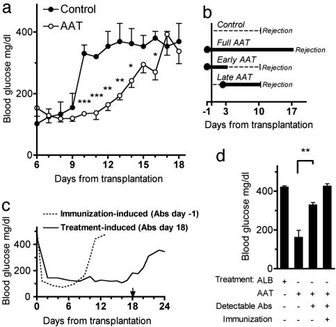
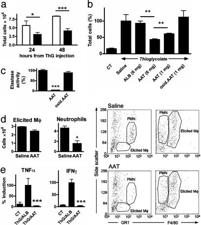
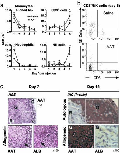
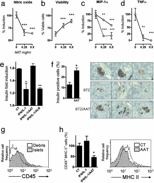
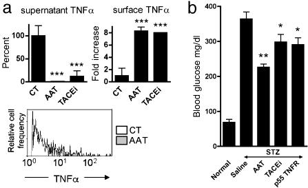
Islet transplantation for type 1 diabetic patients shows promising results with the use of nondiabetogenic immunosuppressive therapy. However, in addition to compromising the immune system of transplant recipients, long-term studies demonstrate that islet viability is impaired. Here, we demonstrate that, in the absence of immunosuppressive agents, monotherapy with clinical-grade human alpha1-antitrypsin (hAAT), the major serum serine-protease inhibitor, prolongs islet graft survival and normoglycemia in transplanted allogeneic diabetic mice, lasting until the development of anti-hAAT antibodies. Compared to untreated or albumin-control-treated graft recipients, which rejected islets at day 10, AAT-treated mice displayed diminished cellular infiltrates and intact intragraft insulin production throughout treatment. Using peritoneal infiltration models, we demonstrate that AAT decreases allogeneic fibroblast-elicited natural-killer-cell influx by 89%, CD3-positive cell influx by 44%, and thioglycolate-elicited neutrophil emigration by 66%. ATT also extended islet viability in mice after streptozotocin-induced beta cell toxicity. In vitro, several islet responses to IL-1beta/IFNgamma stimulation were examined. In the presence of AAT, islets displayed enhanced viability and inducible insulin secretion. Islets also released 36% less nitric oxide and 82% less macrophage inflammatory protein 1 alpha and expressed 63% fewer surface MHC class II molecules. TNFalpha release from IL-1beta/IFNgamma-stimulated islet cells was reduced by 99%, accompanied by an 8-fold increase in the accumulation of membrane TNFalpha on CD45-positive islet cells. In light of the established safety record and the nondiabetogenic potential of AAT, these data suggest that AAT may be beneficial as adjunctive therapy in patients undergoing islet transplantation.


Figures

Fig. 1.
Fig. 1.
Prolonged survival of islet allografts by treatment with AAT. Islets from DBA/2 mice (H-2d) were transplanted under the renal capsule of STZ-induced hyperglycemic C57BL/6 mice (H-2b). (a) Glucose levels from days 6-18. Control consists of graft recipients that were untreated (n = 3) or treated every 3 days (from day-1) with human albumin (6 mg, n = 3). Prolonged islet graft survival is observed in mice treated every 3 days (from day-1) with hAAT (2 mg, n = 10). *, P < 0.05; **, P < 0.01; ***, P < 0.001 between glucose levels on the same day. (b) Treatment protocols. Control and full AAT treatments are represented in a. Shown in b are AAT only on days -1, 1, and 3 (2 mg, n = 3; Early AAT) and AAT from day 2 and every 2 days thereafter (Late AAT) (2 mg, n = 3). The day that glucose levels exceed 300 mg/dl is indicated as Rejection. (c) Effect of mouse anti-hAAT antibodies. The dashed line indicates posttransplantation glucose levels of a mouse (1 representative of 3) under the full AAT treatment protocol (see a and b) that was immunized by prior administrations of hAAT. The solid line indicates glucose levels of a nonimmunized mouse (1 representative of 10) treated under the full AAT treatment protocol. The arrow indicates the day of detection of treatment-induced anti-hAAT antibodies. (d) Comparison of day 15 posttransplantation glucose levels in mice that were under full treatment protocol with albumin (ALB) (n = 3) or AAT (nonimmunized, n = 10; immunized, n = 3). Of the AAT-alone-treated group, antibodies were detected in 3 of 3 immunized mice and in 6 of 10 nonimmunized mice. **, P = 0.005 between mice that produced antibodies and mice that did not.
Fig. 2.
Fig. 2.
Effect of AAT on ThG-elicited peritoneal cellular infiltrates. Mice were administered saline, albumin (ALB), AAT, or oxidized AAT (oxid.ATT), followed by either saline or ThG (3% wt/vol, n = 3 per group). Peritoneal lavage was performed on separate groups after 24 and 48 h. (a) Total cell population of lavaged cells of saline-(open bars) or AAT-treated (5 mg, black bars) ThG-injected mice. *, P < 0.05; ***, P < 0.001. (b) Percent cell population from saline-treated mice at 48 h. **, P < 0.01. CT, control. (c) Oxidation of AAT. AAT was subjected to oxidative radicals, and loss of activity was assessed in an elastase assay. Activity of elastase in the absence of native AAT was set at 100%, and the percentage of activity in the presence of native and oxidized AAT was calculated (n = 3). ***, P < 0.001. (d) Identification of elicited macrophages (Mô) and neutrophils. Peritoneal infiltrates from 48-h lavages of ALB-(6 mg) and AAT-treated (6 mg) ThG-injected mice were identified by FACS analysis. Macrophages and neutrophils were identified on the basis of F4/80 and Gr1 vs. side-scatter flow cytometry profiles. (Left) Quantified FACS results (n = 3). (Right) FACS analysis, representative graphs (n = 3). PMN, polymorphonuclear leukocyte. (e) Production of TNFα by cultured splenocytes from ThG-injected mice. Splenocytes were harvested and washed 48 h after i.p. injection of 1 ml ThG or saline (CT) in mice that were pretreated with albumin (ThG/ALB, 5 mg) or AAT (ThG/AAT, 5 mg). After 48 h of culture in the absence of inducers, levels of secreted TNFα and IFNγ were determined. Mean ± SEM from ThG/ALB group (n = 3 per group).
Fig. 3.
Fig. 3.
Effect of AAT on MHC-incompatible fibroblast-elicited peritoneal cellular infiltrates. Mice (C57BL/6; H-2b) were administered saline or AAT (1 mg) followed by NIH 3T3 cells (1 × 10⁷ cells; H-2d). Peritoneal lavage was performed daily on days 1-5, and cell subpopulations were identified by FACS analysis (n = 3 per treatment). (a) Cell numbers. The number of cells in each subpopulation was calculated from the percentages obtained by FACS analysis and total number of cells in the infiltrate. *, P < 0.05; **, P < 0.01 between cell numbers on the same day. (b) Representative FACS analysis. (c) Effect of AAT on intensity and function of infiltrate elicited by islet allograft. (Left) Hematoxylin and eosin (H&E) staining of day-7 islet allografts. A section of AAT-treated islet graft (in white frame) is compared with an equivalent section from an ALB-treated diabetic recipient mouse (full AAT treatment protocol, see Fig. 1a). The arrow indicates the border between the islet and surrounding infiltrate. (Right) Immunohistochemistry for insulin in islet grafts on day 15. A section of autologous islet graft (in white frame) represents intact transplanted islets and is compared with equivalent sections of allografts of AAT- and ALB-treated recipient mice. R, renal parenchyma; G, graft; C, renal capsule.
Fig. 4.
Fig. 4.
Effect of AAT on islet responses. (a-d) Islets from C57BL/6 mice were cultured (100 islets per well, in duplicate). AAT was incubated at the indicated concentrations for 1 h before the addition of IFNγ (5 ng/ml) and IL-1β (10 ng/ml). Seventy-two hours later, supernatants were collected, and islet viability was assessed. Islet responses in the absence of AAT were set at 100%. The data are combined from three individual experiments. **, P < 0.01; ***, P < 0.001 between AAT-treated and untreated islets. The dashed line represents islets incubated at 1/30th the concentration of IFNγ/IL-1β. (e) Insulin-induction assay. Islets were incubated (20 islets per well, in triplicate) in the presence of AAT (0.5 mg/ml) or ALB (0.5 mg/ml) 1 h before the addition of IFNγ (5 ng/ml) and IL-1β (10 ng/ml). Twenty-four hours later, islets were transferred to a 3 mM or 20 mM glucose solution for 30 min, and insulin levels were measured. The vertical axis represents the ratio between insulin levels at both glucose concentrations. *, P < 0.05 between AAT-treated and ALB-treated islets. (f) In vivo STZ-induced beta cell toxicity. C57BL/6 mice were injected with AAT (5 mg) or saline, 1 day before, on the same day as, and 1 day after injection of STZ (225 mg/kg of body weight) or saline (n = 3 per group). Forty-eight hours later, pancreata were removed, and insulin-containing cells were identified by immunohistochemistry. (Left) Mean ± SEM percent change of insulin-containing cells, as determined from images of two islets per pancreas. (Right) Each image depicts a representative islet from one pancreas (n = 6 per treatment group). *, P < 0.05. (g) Cellular content of islets. Freshly isolated islets (100 islets, in triplicate) and residual nonislet pancreatic debris were dissociated into single-cell suspensions and stained for FACS analysis with anti-CD45-APC or isotype control antibody. (h) MHC class II expression. Islets from C57BL/6 mice were cultured (100 islets per well, in duplicate) in the presence of AAT (0.5 mg/ml) 1 h before the addition of IFNγ (5 ng/ml) and IL-1β (10 ng/ml). Twenty-four hours later, islets were dissociated into single-cell suspensions and double-stained for FACS analysis with anti-CD45-APC and anti-MHCII-PE or isotype control antibodies. (Left) Mean ± SEM percent change from control (CT) unstimulated islets. *, P < 0.05 between AAT-treated and untreated islets. (Right) Representative FACS analysis; events are gated for CD45.
Fig. 5.
Fig. 5.
Effect of AAT on TNFα in islets. (a) Islets from C57BL/6 mice were cultured (100 islets per well, in triplicate) in the presence of AAT (0.5 mg/ml) or TACE inhibitor (10 mM) 1 h before stimulation by IFNγ (5 ng/ml) and IL-1β (10 ng/ml). (Left) Mean (±SEM) change in TNFα levels in supernatants after 72 h of incubation. (Right) Mean (±SEM) fold change in membrane TNFα on islet cells after 5 h of incubation. ***, P < 0.001 compared with control (CT) levels in the absence of AAT. (Lower) Representative FACS analysis of membrane TNFα on stimulated islet cells in the absence (open area) or presence (shaded area) of AAT. Events are gated for CD45. (b) In vivo STZ-induced beta cell toxicity. C57BL/6 mice were injected with saline (n = 3), AAT (5 mg, n = 3) or soluble p55 TNF receptor (p55 TNFR, 1 mg/kg of body weight, n = 3) or administered oral TACE inhibitor (TACEi, 60 mg/kg of body weight, n = 6) 1 day before injection of STZ (225 mg/kg of body weight, i.p.). Subsequently, AAT and soluble TNF receptor were injected daily, and TACE inhibitor was administered twice a day. At 48 h, mean (±SEM) glucose levels are compared with those of normal littermates (n = 3). *, P < 0.05; **, P < 0.01, compared with saline-treated, STZ-injected mice.

Comment in

  • Saving islets from allograft rejection.
    Strom TB. Strom TB. Proc Natl Acad Sci U S A. 2005 Sep 6;102(36):12651-2. doi: 10.1073/pnas.0506079102. Epub 2005 Aug 29. Proc Natl Acad Sci U S A. 2005. PMID: 16129823 Free PMC article. No abstract available.

Similar articles

Cited by

References

    1. Sato, T., Inagaki, A., Uchida, K., Ueki, T., Goto, N., Matsuoka, S., Katayama, A., Haba, T., Tominaga, Y., Okajima, Y., et al. (2003) Transplantation 76, 1320-1326. - PubMed
    1. Ricordi, C. & Strom, T. B. (2004) Nat. Rev. Immunol. 4, 259-268. - PubMed
    1. Couzin, J. (2004) Science 306, 34-37. - PubMed
    1. Kobayashi, N., Okitsu, T., Lakey, J. R. & Tanaka, N. (2004) J. Artif. Organs 7, 1-8. - PubMed
    1. Breit, S. N., Wakefield, D., Robinson, J. P., Luckhurst, E., Clark, P. & Penny, R. (1985) Clin. Immunol. Immunopathol. 35, 363-380. - PubMed

Publication types

MeSH terms