Comparative Study
. 2005 Oct;94(4):2763-71.
doi: 10.1152/jn.00351.2005. Epub 2005 Aug 10.

Cholecystokinin octapeptide increases spontaneous glutamatergic synaptic transmission to neurons of the nucleus tractus solitarius centralis

Affiliations
Comparative Study

Cholecystokinin octapeptide increases spontaneous glutamatergic synaptic transmission to neurons of the nucleus tractus solitarius centralis

V Baptista et al. J Neurophysiol. 2005 Oct.

Abstract

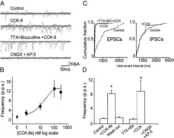
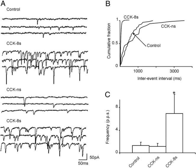
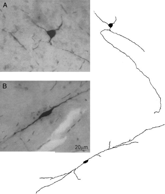
Cholecystokinin (CCK) is released from enteroendocrine cells after ingestion of nutrients and induces multiple effects along the gastrointestinal tract, including gastric relaxation and short-term satiety. We used whole cell patch-clamp and immunohistochemical techniques in rat brain stem slices to characterize the effects of CCK. In 45% of the neurons of nucleus tractus solitarius subnucleus centralis (cNTS), perfusion with the sulfated form of CCK (CCK-8s) increased the frequency of spontaneous excitatory currents (sEPSCs) in a concentration-dependent manner (1-300 nM). The threshold for the CCK-8s excitatory effect was 1 nM, the EC(50) was 20 nM, and E(max) was 100 nM. The excitatory effects of CCK-8s were still present when the slices were preincubated with tetrodotoxin or bicuculline or when the recordings were conducted with Cs(+) electrodes. Pretreatment with the CCK-A receptor antagonist, lorglumide (1 microM), antagonized the effects of CCK-8s, whereas perfusion with the CCK-B preferring agonist CCK-8 nonsulfated (CCK-ns, 1 microM) did not affect the frequency of sEPSCs. Similarly, pretreatment with the CCK-B receptor antagonist, triglumide (1 microM), did not prevent the actions of CCK-8s. Although the majority (i.e., 76%) of CCK-8s unresponsive cNTS neurons had a bipolar somata shape and were TH-IR negative, no differences were found in either the morphological or the neurochemical phenotype of cNTS neurons responsive to CCK-8s. Our results suggest that the excitatory effects of CCK-8s on terminals impinging on a subpopulation of cNTS neurons are mediated by CCK-A receptors; these responsive neurons, however, do not have morphological or neurochemical characteristics that automatically distinguish them from nonresponsive neurons.


Figures

FIG. 1
FIG. 1
A: representative traces showing spontaneous excitatory postsynaptic currents (EPSCs) in control conditions (top); perfusion with 100 nM of sulfated cholecystokinin (CCK-8s) increased the frequency of spontaneous EPSCs but did not alter the frequency of spontaneous inhibitory postsynaptic current (IPSC) (middle); the effects of CCK-8s were still present after tetrodotoxin (TTX; 1 μM) and bicuculline (50 μM) pretreatment (middle, low) but not CNQX (10 μM) or AP-5 (30 μM) (bottom). B: concentration–response curve for the CCK-8s–induced increase in spontaneous EPSC (n = 5 for each data point). C: computer-generated graphics from the same neuron as in A showing that CCK-8s increased the frequency of EPSCs but not IPSCs. D: graphics summarizing the effects of 100 nM CCK-8s on frequency of spontaneous (left) and miniature (right) EPSCs. *P < 0.05 vs. control.
FIG. 2
FIG. 2
A: representative traces showing spontaneous EPSCs in control conditions (left); after 10-min perfusion with the CCK-B receptor antagonist, triglumide (1 μM), perfusion with 100 nM CCK-8s still increased the frequency of spontaneous EPSCs (middle); the effects of CCK-8s, however, were antagonized by 10-min perfusion with the CCK-A receptor antagonist, lorglumide (1 μM; right). Traces are from the same nucleus tractus solitarius subnucleus centralis (cNTS) neuron. Holding potential = −50 mV. B: graphics summarizing the effects of 100 nM CCK-8s on frequency of spontaneous EPSCs in the presence of the selective CCK-A (right) or CCK-B (left) receptor antagonists. *P < 0.05 vs. control.
FIG. 3
FIG. 3
A: representative traces showing spontaneous EPSCs in control conditions, perfusion with 100 nM CCK-8s increased the frequency of spontaneous EPSCs; after a 10-min washout of CCK-8s, perfusion with the CCK-B preferring agonist, nonsulfated cholecystokinin (CCK-ns; 1 μM) did not increase the frequency of spontaneous EPSCs; however, perfusion with 100 nM CCK-8s immediately after CCK-ns still increased the frequency of spontaneous EPSCs. Holding potential = −50 mV. B: computer-generated graphics from the same neuron as in A showing that CCK-8s, but not CCK-ns, increased the frequency of spontaneous EPSCs. C: graphics summarizing the effects of 100 nM CCK-8s and 1 μM CCK-ns on frequency of spontaneous EPSCs. *P < 0.05 vs. control.
FIG. 4
FIG. 4
A, left: micrograph of a cNTS neuron with multipolar somata shape; right: computer-aided reconstruction of the neuron on left. B, left: micrograph of a cNTS neuron with bipolar somata shape; right: computer-aided reconstruction of the neuron on left.
FIG. 5
FIG. 5
A: schematic diagram depicting the localization of cNTS neurons according to their soma shape (bipolar = square; multipolar = circle) and their response to perfusion with 100 nM CCK-8s (responsive to CCK-8s = white symbol; nonresponsive to CCK-8s = black symbol). For the purpose of intelligibility, not all the neurons have been reported in the graphic. TS, tractus solitarius; AP, area postrema; DMV, dorsal motor nucleus of the vagus; cc, central canal; IV Ventr., fourth ventricle. B: micrographs showing a postrecording immunohistochemical detection in a tyrosine-hydroxylase immunoreactive (TH-IR) positive (left) and in a TH-IR negative (right) cNTS neuron. Scale bar = 10 μm.

Similar articles

Cited by

References

    1. Altschuler SM, Bao X, Bieger D, Hopkins DA, Miselis RR. Viscerotopic representation of the upper alimentary tract in the rat: sensory ganglia and nuclei of the solitary and spinal trigeminal tracts. J Comp Neurol. 1989;283:248–268. - PubMed
    1. Andresen MC, Yang M. Non-NMDA receptors mediate sensory afferent synaptic transmission in medial nucleus tractus solitarius. Am J Physiol Heart Circ Physiol. 1990;259:H1307–H1311. - PubMed
    1. Appleyard SM, Bailey TW, Doyle MW, Jin YH, Smart JL, Low MJ, Andresen MC. Proopiomelanocortin neurons in nucleus tractus solitarius are activated by visceral afferents: regulation by cholecystokinin and opioids. J Neurosci. 2005;25:3578–3585. - PMC - PubMed
    1. Baptista V, Zheng ZL, Coleman FH, Rogers RC, Travagli RA. Characterization of neurons of the nucleus tractus solitarius pars centralis. Brain Res. 2005;1052:139–146. - PMC - PubMed
    1. Barraco R, El-Ridi M, Parizon M, Bradley D. An atlas of the rat subpostremal nucleus tractus solitarius. Brain Res Bull. 1992;29:703–765. - PubMed

Publication types

MeSH terms