Comparative Study
. 2005 Dec;94(6):3938-50.
doi: 10.1152/jn.00340.2005. Epub 2005 Aug 10.

Myomodulin increases Ih and inhibits the NA/K pump to modulate bursting in leech heart interneurons

Affiliations
Comparative Study

Myomodulin increases Ih and inhibits the NA/K pump to modulate bursting in leech heart interneurons

Anne-Elise Tobin et al. J Neurophysiol. 2005 Dec.

Abstract

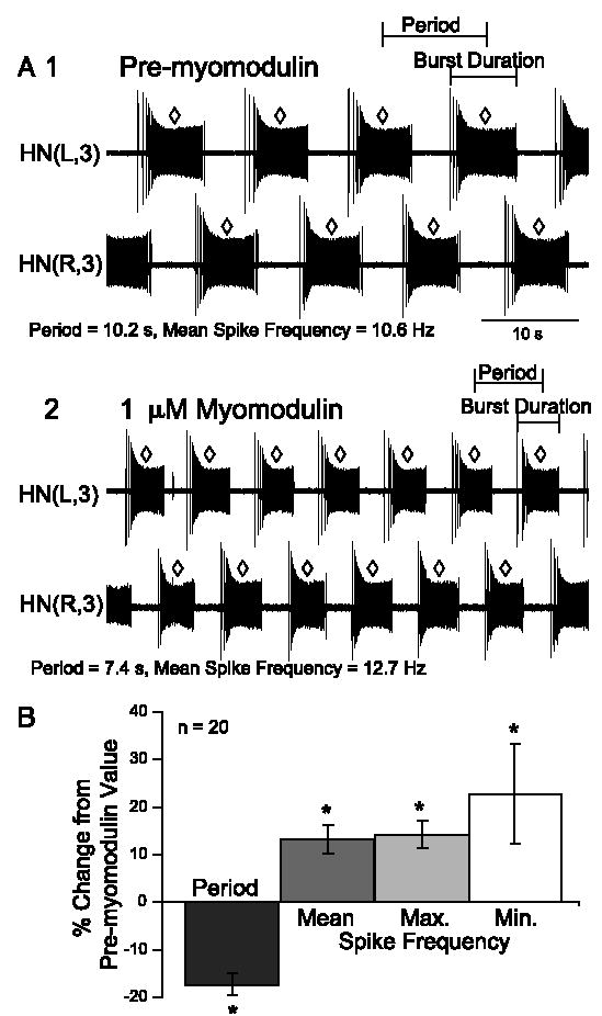
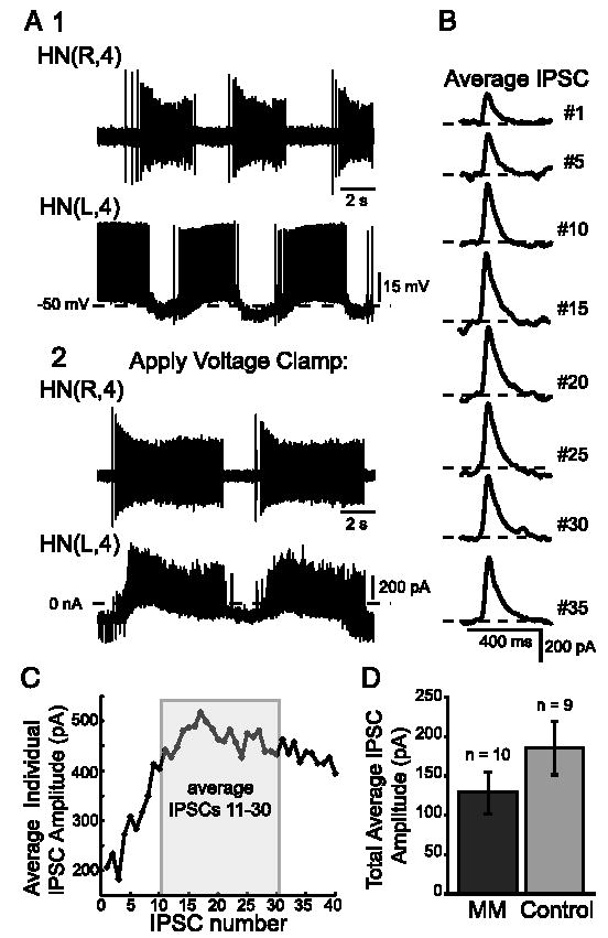
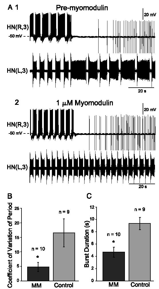
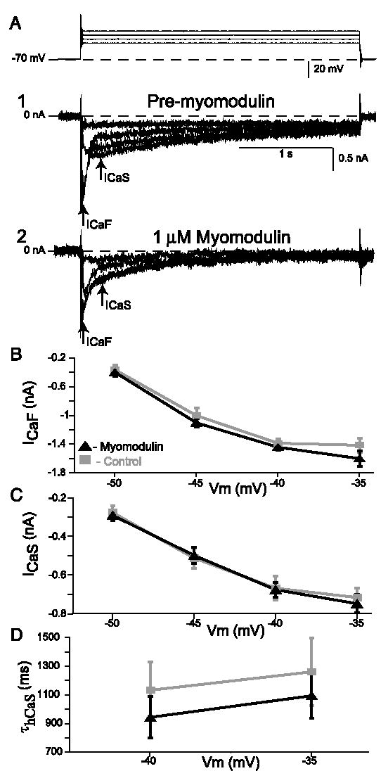
In the medicinal leech, a rhythmically active 14-interneuron network composes the central pattern generator for heartbeat. In two segmental ganglia, bilateral pairs of reciprocally inhibitory heart interneurons (oscillator interneurons) produce a rhythm of alternating bursts of action potentials that paces activity in the pattern-generating network. The neuropeptide myomodulin decreases the period of this bursting and increases the intraburst spike frequency when applied to isolated ganglia containing these oscillator interneurons. Myomodulin also decreases period, increases spike frequency, and increases the robustness of endogenous bursting in synaptically isolated (with bicuculline) oscillator interneurons. In voltage-clamp experiments using hyperpolarizing ramps, we identify an increase in membrane conductance elicited by myomodulin with the properties of a hyperpolarization-activated current. Voltage steps confirm that myomodulin indeed increases the maximum conductance of the hyperpolarization-activated current I(h). In similar experiments using Cs(+) to block I(h), we demonstrate that myomodulin also causes a steady offset in the ramp current that is not associated with an increase in conductance. This current offset is blocked by ouabain, indicating that myomodulin inhibits the Na/K pump. In current-clamp experiments, when I(h) is blocked with Cs(+), myomodulin decreases period and increases spike frequency of alternating bursting in synaptically connected oscillator interneurons, suggesting that inhibiting the Na/K pump modulates these burst characteristics. These observations indicate that myomodulin decreases period and increases spike frequency of endogenous bursting in synaptically isolated oscillator heart interneurons and alternating bursting of reciprocally inhibitory pairs of interneurons, at least in part, by increasing I(h) and by decreasing the Na/K pump.


Figures

Fig. 1
Fig. 1
Myomodulin changes the burst characteristics of oscillator heart interneurons. A: example extracellular recordings from heart interneurons in ganglion 3: HN(L,3) and HN(R,3). Period is measured as the time between median spikes (diamonds) of consecutive bursts. Burst duration is measured as the time from first to last spike in a burst. Before myomodulin application (1), HN(R,3) burst period was 10.2 s and mean intraburst spike frequency was 10.6 Hz. When 1 μM myomodulin was added (2), HN(R,3) period decreased to 7.4 s and mean spike frequency increased to 12.7 Hz. B: myomodulin decreased burst period while increasing mean, maximum, and minimum intraburst spike frequencies. * indicates significant difference from 0.
Fig. 2
Fig. 2
Myomodulin-induced changes in burst properties are correlated with their premyomodulin values. Black diamonds indicate heart interneurons from ganglion 3 [HN(3)], gray squares indicate heart interneurons from ganglion 4 [HN(4)], and white triangles indicate data where ganglion number was not recorded [HN(?)]. A: myomodulin decreased period more for oscillator heart interneurons with larger initial periods. B: myomodulin increased spike frequency more for oscillator heart interneurons with smaller initial mean spike frequencies.
Fig. 3
Fig. 3
Myomodulin does not change the amplitude of spike-mediated synaptic currents. A, 1: extracellular (top) and intracellular (bottom) recordings from a pair of heart interneurons in ganglion 4; 2: left interneuron [HN(L,4); bottom] was voltage-clamped at −50 mV and its voltage-clamp current is shown. B: inhibitory postsynaptic currents (IPSCs) were averaged according to their sequence in each burst. Dashed line indicates zero current. C: average individual IPSC amplitude increased then slightly waned during each burst. Amplitudes of IPSCs 11–30 were averaged to obtain the total average IPSC amplitude for each experiment. D: myomodulin (MM) did not change the total average IPSC amplitude.
Fig. 4
Fig. 4
Myomodulin increases burst robustness in an oscillator heart inter-neuron whose partner is voltage clamped. A: heart interneurons from ganglion 3 burst in alternation. 1: when the right neuron [HN(R,3)] was prevented from bursting by voltage clamp, the left neuron [HN(L,3)], recorded extracellularly, exhibited prolonged, irregular bursts; 2: when 1 μM myomodulin was added, the unclamped neuron maintained regular bursting while the partner neuron was voltage clamped. Robust bursting in the presence of myomodulin (MM) was indicated by a decrease in the coefficient of variation of period (B) and a decrease in burst duration (C).
Fig. 5
Fig. 5
Myomodulin increases burst robustness in synaptically isolated heart interneurons. A, 1: heart interneurons from ganglion 3, HN(R,3) and HN(L,3), burst endogenously when synapses were blocked by 0.5 mM bicu-culline. Irregular bursting sometimes changed to tonic spiking for many seconds; 2: when 1 μM myomodulin was added, bursting became regular and bursts became more discrete. Robust bursting in myomodulin (MM) was indicated by a decrease in the coefficient of variation of period (B) and decrease in burst duration (C), compared with the pre-myomodulin (pre-MM) condition.
Fig. 6
Fig. 6
Myomodulin increases membrane conductance during hyperpolarizing voltage ramps. A: average voltage-clamp current for myomodulin application (black) and control (gray), from 9 and 8 experiments each. Myomodulin increased conductance, as indicated by the steeper slope of the clamp current. Inset: command voltage. B and C: subtracting the average voltage-clamp current with myomodulin application from that with control yielded the difference current. Difference current corresponding to hyperpolarization is indicated by downward arrows; current corresponding to repolarization is indicated by upward arrows. C: myomodulin elicited a current that was slowly activated by hyperpolarization, as shown by plotting the difference current vs. voltage.
Fig. 7
Fig. 7
Myomodulin does not change K currents elicited during hyperpolarizing voltage-clamp steps. A: example voltage (top) and current traces (1, 2) during a voltage-clamp protocol to elicit K currents. Amplitude of K currents was measured as the difference between the current 10–20 ms before pulse onset (left arrow) and the steady-state current (right arrow). 1, 2: example voltage-clamp current before (1) and after (2) application of 1 μM myomodulin. B: myomodulin (black triangles) does not change the amplitude of steady-state K currents, compared with control (gray squares).
Fig. 8
Fig. 8
Myomodulin increases maximal conductance of Ih elicited by voltage-clamp steps in 0 Ca2+, 1.8 mM Mn2+ saline. A: example voltage (top) and current traces (1, 2) during a voltage-clamp protocol to elicit Ih. Amplitude of Ih was measured as the difference between the current 75–85 ms after pulse onset (left arrow) and the steady-state current (right arrow). 1, 2: example voltage-clamp currents before (1) and after (2) application of 1 μM myomodu-lin. B: myomodulin (black triangles) increased Ih conductance for steps to −80 and −100 mV, compared with control (gray squares). C: change in Ih conductance from pretreatment measurements was significantly larger in myomodulin than in control experiments for all voltage steps.
Fig. 9
Fig. 9
In 0 Ca2+, 1.8 mM Mn2+ saline with 2 mM Cs+, myomodulin causes an offset in voltage-clamp current during hyperpolarizing voltage ramps that is blocked by ouabain. A: average voltage-clamp current for myomodulin application (black) and control (gray), from 10 and 9 experiments, respectively. Slopes of the current during the hyperpolarizing and depolarizing phases of the ramp were the same for the myomodulin application and control condition, indicating similar conductance. Holding and ramp current during the myomodulin application were offset from the control condition. Inset: command voltage. B: in experiments where ouabain was present in 0 Ca2+ saline with 2 mM Cs+, there was no myomodulin-induced current offset. There was no offset in holding or ramp current between myomodulin and control conditions; averages from 11 and 9 experiments, respectively.
Fig. 10
Fig. 10
Myomodulin-induced changes of burst characteristics in Cs+. A, 1: : a heart interneuron from ganglion 3, HN(R,3), burst when Ih was blocked by 2 mM Cs+; 2: when 1 μM myomodulin was added, period decreased and mean spike frequency increased. B: average change in period and mean spike frequency. * indicates significant difference from 0.
Fig. 11
Fig. 11
Myomodulin does not change low-threshold Ca currents. A: example voltage (top) and current traces (1, 2) from a voltage-clamp experiment to measure slow and fast Ca currents. Ca2+ concentration was elevated to 5 mM to enhance Ca currents. Amplitude of the fast Ca current (ICaF) was measured as the peak Ca current within the first 200 ms of the step (left arrow). Amplitude of the slow Ca current (ICaS) was measured as the subsequent peak Ca current within the first 500 ms of the step (right arrow). ICaS inactivation time constant (τhCaS) was measured as the longest time constant in a 4 exponential fit of the Ca current waveform. 1, 2: example voltage-clamp currents before (1) and after (2) application of 1 μM myomodulin. B: myomodulin does not change peak ICaF amplitude (for steps to −50, −45, −40, and −35 mV, respectively: control, n = 9, 9, 9, 5; myomodulin, n = 9, 9, 8, 6). C: myomodulin does not change peak ICaS amplitude (control, n = 10; myomodulin, n = 9). D: myomodulin does not change τhCaS (for steps to −40 and −35 mV, respectively: control, n = 9, 9; myomodulin, n = 9, 10).

Similar articles

Cited by

References

    1. Aizman O, Brismar H, Uhlen P, Zettergren E, Levey AI, Forssberg H, Greengard P, Aperia A. Anatomical and physiological evidence for D1 and D2 dopamine receptor colocalization in neostriatal neurons. Nat Neurosci. 2000;3:226–230. - PubMed
    1. Angstadt JD, Calabrese RL. A hyperpolarization-activated inward current in heart interneurons of the medicinal leech. J Neurosci. 1989;9:2846–2857. - PMC - PubMed
    1. Angstadt JD, Calabrese RL. Calcium currents and graded synaptic transmission between heart interneurons of the leech. J Neurosci. 1991;11:746–759. - PMC - PubMed
    1. Angstadt JD, Friesen WO. Synchronized oscillatory activity in leech neurons induced by calcium channel blockers. J Neurophysiol. 1991;66:1858–1873. - PubMed
    1. Arbas EA. Rate modification in the heartbeat central pattern generator of the medicinal leech. J Comp Physiol A Sens Neural Behav Physiol. 1984;155:783–794.

Publication types

MeSH terms

LinkOut - more resources