. 2005 Oct;16(10):4992-5003.
doi: 10.1091/mbc.e05-04-0310. Epub 2005 Aug 10.

Dual interaction of JAM-C with JAM-B and alpha(M)beta2 integrin: function in junctional complexes and leukocyte adhesion

Affiliations

Dual interaction of JAM-C with JAM-B and alpha(M)beta2 integrin: function in junctional complexes and leukocyte adhesion

Chrystelle Lamagna et al. Mol Biol Cell. 2005 Oct.

Abstract

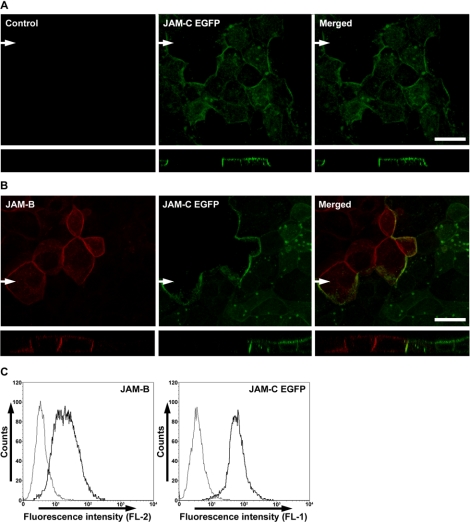
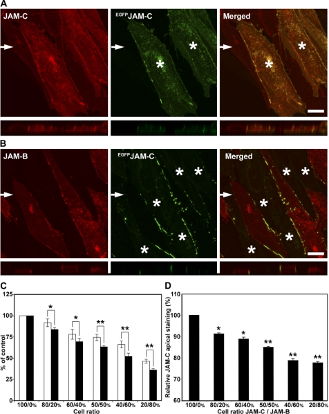
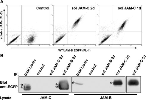
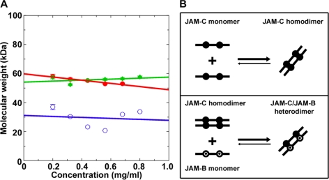
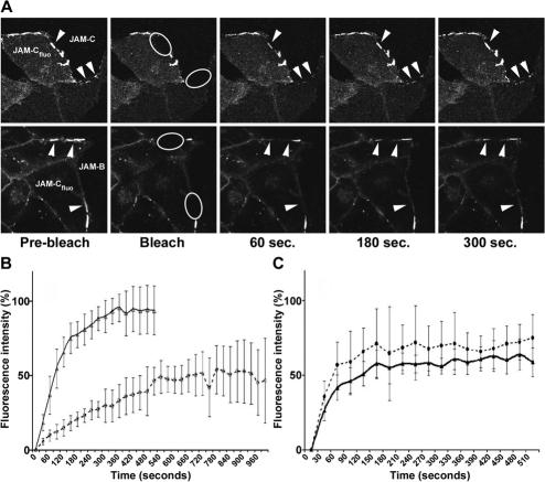
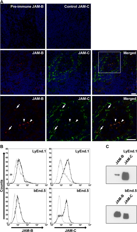
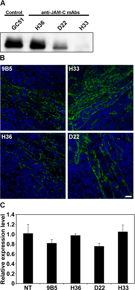
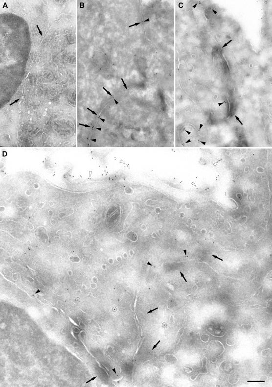
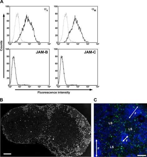
The junctional adhesion molecules (JAMs) have been recently described as interendothelial junctional molecules and as integrin ligands. Here we show that JAM-B and JAM-C undergo heterophilic interaction in cell-cell contacts and that JAM-C is recruited and stabilized in junctional complexes by JAM-B. In addition, soluble JAM-B dissociates soluble JAM-C homodimers to form JAM-B/JAM-C heterodimers. This suggests that the affinity of JAM-C monomers to form dimers is higher for JAM-B than for JAM-C. Using antibodies against JAM-C, the formation of JAM-B/JAM-C heterodimers can be abolished. This liberates JAM-C from its vascular binding partner JAM-B and makes it available on the apical side of vessels for interaction with its leukocyte counter-receptor alpha(M)beta2 integrin. We demonstrate that the modulation of JAM-C localization in junctional complexes is a new regulatory mechanism for alpha(M)beta2-dependent adhesion of leukocytes.


Figures

Figure 1.
Figure 1.
JAM-C is recruited at cell-cell contacts by JAM-B. FLAG-JAM-B- and JAM-C-EGFP-transfected MDCK cells were mixed and the localization of the molecules was visualized by immunofluorescent labeling. (A) Negative control for JAM-B staining with polyclonal rabbit antibody and anti-rabbit Texas red probe. (B) Plane view of stacked series of pictures (top panel), JAM-C-EGFP is enriched in JAM-C/JAM-B intercellular contacts. On the Z-axis analysis (bottom panel), JAM-C appears to be localized in basolateral contacts when engaged heterophilically with JAM-B contacts. Arrows indicate the Z-axis. Scale bar, 20 μm. (C) Surface expression of JAM-B and JAM-C EGFP on MDCK cells used in A and B. The thin line represents the negative control obtained by omitting the primary antibody; bold line represents surface expression of JAM-B or JAM-C EGFP, as indicated.
Figure 2.
Figure 2.
Recruitment of JAM-C at cell-cell contacts depletes the apical pool of the protein. (A) CHO cells transfected with EGFPJAM-C (green) were mixed with JAM-C full-length (red) or (B) JAM-B-expressing cells (red) and stained with polyclonal antibodies against murine JAM-B or JAM-C. The plane view of stacked series of images and Z-axis reconstitution show that EGFPJAM-C is enriched in JAM-C/JAM-B intercellular contacts as compared with JAM-C/JAM-C intercellular contacts. Arrows indicate the Z-axis. Scale bar, 20 μm. (C) Quantification by ELISA of EGFPJAM-C expressed on the apical cell surface. EGFPJAM-C-transfected CHO cells were mixed with JAM-C (white columns) or JAM-B-expressing cells (black columns) at indicated cell ratios. The protein EGFPJAM-C at the apical cell surface was detected with an anti-JAM-C polyclonal antibody. (D) Same as B, but signals were normalized to the ones obtained with mixed monolayers of JAM-C-expressing cells with nontransfected cells. As shown, the relative JAM-C signal detected at the apical cell surface is decreased when the percentage of JAM-B-positive cells is increased. Each bar represents the mean value ± SEM (n = 6) of one representative experiment observed in at least three separate experiments. (* p < 0.05, ** p < 0.01).
Figure 3.
Figure 3.
JAM-C interacts heterophilically with JAM-B through its V domain, and monomeric JAM-B dissociates JAM-C homodimers to form heterodimers. (A) JAM-B-EGFP-transfected MDCK cells were mixed with nontransfected cells and stained with soluble JAM-C consisting in the two extracellular domains (2d) or the membrane distal V domain (1d) alone. Dot plots represent the EGFP fluorescence intensity (FL1) and the fluorescence intensity due to binding of soluble molecules (FL2). The soluble JAM-C 2d as well as the soluble JAM-C 1d binds to JAM-B-EGFP-transfected cells. No binding was observed on nontransfected cells as depicted by the absence of FL2 signal on EGFP-negative cells. Negative control obtained by omitting primary antibody against the flag-tag is shown. (B) Cell lysates obtained from JAM-C-EGFP- or JAM-B-EGFP-transfected MDCK cells were precipitated with beads coupled to soluble JAM-B or soluble JAM-C. Western blots were revealed using anti-EGFP antibody. Soluble JAM-C 2d, as well as soluble JAM-C 1d, and soluble JAM-B are able to pull-down JAM-B-EGFP and JAM-C-EGFP, respectively. Control of loading was obtained using M2 anti-FLAG antibody (unpublished data).
Figure 4.
Figure 4.
Monomeric JAM-B dissociates JAM-C homodimers to form JAM-B/JAM-C heterodimers. (A) Representative plots of apparent whole-cell weight-average molecular weight (Mw) against sample concentration (mg/ml) for absorbance data at 280 nm and 12 000 rpm. Open blue circles are for JAM-B, red circles are for JAM-C, and green diamonds are for an equimolar JAM-B/JAM-C mixture. Fitted curves are for a linear regression of weight with concentration as appropriate for a dimerization process (Ikemizu et al., 2000). (B) Schematic representation of JAM-B/JAM-C heterodimerization. In solution, JAM-C tends to form homodimers (top panel). In the presence of JAM-B molecule, JAM-B/JAM-C heterodimers are preferentially formed. This interpretation does not account for parallel or antiparallel heterodimerization.
Figure 5.
Figure 5.
Dynamic of JAM-C junctional recruitment by JAM-B in CHO-transfected cells. (A) EGFPJAM-C CHO cells were mixed with JAM-B CHO cells at a ratio 1:1. FRAP experiments of EGFPJAM-C engaged in homophilic interaction (top panel) or heterophilic interaction (bottom panel) were performed. Regions of interest for bleaching were drawn on contacts between EGFPJAM-C-expressing cells and nonfluorescent cells (ellipses). Arrowheads underline clusters of EGFPJAM-C in which the recovery occurred. Elapsed time after photobleaching is indicated. Scale bars, 10 μm. (B) Fractional recovery curves obtained for EGFPJAM-C/JAM-C (plain curve, open triangles) or EGFPJAM-C/JAM-B (dashed curve, open circles) junctional complexes. Recovered fluorescence is expressed as function of time elapsed after photobleaching (time 0). (C) Fractional recovery curves obtained for PECAM-EGFP/JAM-C (plain curve, filled triangles) or PECAM-EGFP/JAM-B (dashed curve, filled circles) cell-cell contacts are shown. No significant differences can be observed between the two recovery curves indicating that the differences observed in B are due to trans-interaction between EGFPJAM-C and its binding partners JAM-B or JAM-C. The curves are mean values ± SD obtained from at least four independent experiments and three to four regions of interest analyzed for each acquisition.
Figure 6.
Figure 6.
JAM-C is predominantly expressed by lymphatic vessels, whereas JAM-B is prominent in vascular endothelial cells. (A) Double staining for JAM-B (red) and JAM-C (green) performed on murine peripheral lymph node sections shows that JAM-C is highly expressed on lymphatic sinuses (arrows), whereas JAM-B is strongly expressed by HEVs. Higher magnification (bottom panel) shows that JAM-B and JAM-C are both expressed on high endothelial venules where they partially colocalize (arrowheads). Negative control obtained with preimmune rabbit serum against JAM-B or by omitting primary antibody against JAM-C are shown. Scale bar, 20 μm. (B) The expression levels of JAM-B and JAM-C molecules by the lymphangioma cell line LyEnd.1 (Supplementary Figure 4) and the endothelioma cell line bEnd.5 were analyzed by FACS. JAM-C expression is predominant in LyEnd.1, whereas JAM-B is preferentially expressed by endothelial cells from blood origin. (C) JAM-B and JAM-C expressions by LyEnd.1 (top panel) and bEnd.5 (bottom panel) were analyzed by Western blot and enforced the results observed by FACS.
Figure 7.
Figure 7.
Anti-JAM-C mAb blocks JAM-B/JAM-C interaction and modulates JAM-C localization. (A) Cell lysates obtained from JAM-B-EGFP-transfected MDCK cells were precipitated with beads coupled to soluble JAM-C in the presence of irrelevant anti-PECAM antibody (GC51) or antibodies directed against JAM-C, as indicated. The presence of immunoprecipitated material was revealed by immunoblotting with antibody directed against EGFP. The anti-JAM-C antibody H33 abolishes JAM-C/JAM-B interaction. (B) Sections of lymph nodes harvested from mice treated with the indicated antibodies were stained with an anti-JAM-C polyclonal antibody (green) and for nuclei (blue). The H33 antibody, which affects JAM-B/JAM-C interaction, induces redistribution of JAM-C as compared with the nonblocking anti-JAM-C antibody H36 or the isotype-matched control (9B5). This was observed on sections obtained from three different mice. One representative picture is shown. Scale bar, 20 μm. (C) Relative expression levels of JAM-C mRNA in lymph nodes from antibody-treated mice were quantified by real-time PCR. Treatment of animals with anti-JAM-C antibodies, and specifically H33 antibody, does not affect JAM-C expression by endothelial cells. Each bar represents the mean ± SEM of three mice. One representative experiment out of three.
Figure 8.
Figure 8.
Anti-JAM-C antibody H33 redistributes JAM-C away from junctional regions of endothelial cells in vivo. Ultrathin frozen sections of lymphatic regions in lymph nodes from control and anti-JAM-C antibody-treated mice were immunolabeled for JAM-C. (A) After incubation of a section of a control lymph node with a preimmune rabbit serum and a goat serum against rabbit IgG conjugated with gold particles, no staining was observed along the apposed, lateral membranes (arrows) of endothelial cells. (B-D) Labeling of lymph node sections with immune serum against JAM-C resulted in a specific staining of lateral membranes, shown here in an apical (top) to basal orientation (bottom), in both control and H33-treated lymph nodes. (B) In control lymph node, most junctional regions (arrows) were immunolabeled for JAM-C (arrowheads). (C) In a lymph node from a mouse treated with the H33 antibody, JAM-C (arrowheads) was usually not detected at junctional regions (arrows), but was evident in nearby, nonjunctional regions of the same lateral membrane (arrowheads). (D) JAM-C was immunodetected in the cytoplasm (circled), at the apical membrane of endothelial cells (open arrow heads), and at lateral cell-to-cell interfaces (solid arrow heads). Bars, 200 nm in A-C, 400 nm in D.
Figure 9.
Figure 9.
Leukocyte adhesion to frozen section of lymph nodes (Stamper-Woodruff assay). (A) Surface expression of the indicated adhesion molecules by WEHI78/24 monocytoid cell line were detected by flow cytometry using monoclonal antibodies against α4 integrin (PS/2), αM integrin (M1/70), or polyclonal sera against murine JAM-B and JAM-C. Plain profiles show the surface expression, whereas dashed profiles show the appropriate isotype matched controls for rat monoclonal antibodies or preimmune rabbit sera. (B) Representative image of lymph node section to which the monocytoid cell line labeled with calcein adhered for 30 min at 37°C. Most of the fluorescent monocytes adhered to subcapsular areas typical of lymphatic sinuses. Scale bar, 250 μm. (C) Higher magnification of lymph node section (nuclei stained in blue) after adhesion of calcein-labeled monocytoid cells (green). F, follicles; LS, lymphatic sinuses. Scale bar, 100 μm.
Figure 10.
Figure 10.
Treatment of mice with antibodies to JAM-C increases monocyte adhesion to lymph node sections in Stamper and Woodruff assays. (A) Stamper and Woodruff assay was performed on lymph node sections obtained from nontreated animals. WEHI78/24 cells were incubated in the presence or absence of indicated anti-JAM-C antibodies. In these conditions anti-JAM-C antibodies do not affect the adhesion of monocytoid cells to lymph node sections. (B) Stamper and Woodruff adhesion assay was done on lymph node sections from mice treated with H36, D22, H33, or isotype-matched control antibodies. As shown, H33 antibody increases the adhesion of monocytoid cells when administrated to mice. Experiments were done in the presence of blocking antibodies against α4 integrin (PS/2, white columns), against αM integrin (M1/70, dashed columns) or isotype-matched control antibody (black columns). Data shown are the mean ± SEM of the number of adhering cells/mm2 found on eight sections per lymph nodes in three animals per condition. * p < 0.05.

Similar articles

Cited by

References

    1. Altamirano, M. M., Woolfson, A., Donda, A., Shamshiev, A., Briseno-Roa, L., Foster, N. W., Veprintsev, D. B., De Libero, G., Fersht, A. R., and Milstein, C. (2001). Ligand-independent assembly of recombinant human CD1 by using oxidative refolding chromatography. Proc. Natl. Acad. Sci. USA 98, 3288-3293. - PMC - PubMed
    1. Arrate, M. P., Rodriguez, J. M., Tran, T. M., Brock, T. A., and Cunningham, S. A. (2001). Cloning of human junctional adhesion molecule 3 (JAM3) and its identification as the JAM2 counter-receptor. J. Biol. Chem. 276, 45826-45832. - PubMed
    1. Aurrand-Lions, M., Duncan, L., Ballestrem, C., and Imhof, B. A. (2001a). JAM-2, a novel Ig superfamily molecule, expressed by endothelial and lymphatic cells. J. Biol. Chem. 276, 2733-2741. - PubMed
    1. Aurrand-Lions, M., Johnson-Leger, C., Wong, C., Du Pasquier, L., and Imhof, B. A. (2001b). Heterogeneity of endothelial junctions is reflected by differential expression and specific subcellular localization of the three JAM family members. Blood 98, 3699-3707. - PubMed
    1. Aurrand-Lions, M., Lamagna, C., Dangerfield, J. P., Wang, S., Herrera, P., Nourshargh, S., and Imhof, B. A. (2005). Junctional adhesion molecule-C regulates the early influx of leukocytes into tissues during inflammation. J. Immunol. 174, 6406-6415. - PubMed

Publication types

MeSH terms