. 2005 Aug 10;25(32):7299-308.
doi: 10.1523/JNEUROSCI.1573-05.2005.

Phosphorylation of synapsin I by cAMP-dependent protein kinase controls synaptic vesicle dynamics in developing neurons

Affiliations

Phosphorylation of synapsin I by cAMP-dependent protein kinase controls synaptic vesicle dynamics in developing neurons

Dario Bonanomi et al. J Neurosci. .

Abstract

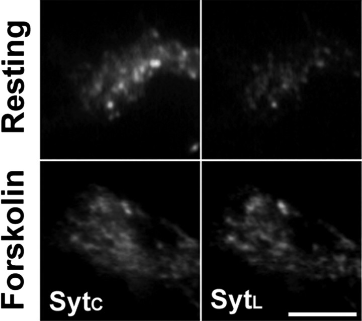
In developing neurons, synaptic vesicles (SVs) undergo cycles of exo-endocytosis along isolated axons. However, it is currently unknown whether SV exocytosis is regulated before synaptogenesis. Here, we show that cAMP-dependent pathways affect SV distribution and recycling in the axonal growth cone and that these effects are mediated by the SV-associated phosphoprotein synapsin I. The presence of synapsin I on SVs is necessary for the correct localization of the vesicles in the central portion of the growth cone. Phosphorylation of synapsin I by cAMP-dependent protein kinase (protein kinase A) causes the dissociation of the protein from the SV membrane, allowing diffusion of the vesicles to the periphery of the growth cone and enhancing their rate of recycling. These results provide new clues as to the bases of the well known activity of synapsin I in synapse maturation and indicate that molecular mechanisms similar to those operating at mature nerve terminals are active in developing neurons to regulate the SV life cycle before synaptogenesis.


Figures

Figure 1.
Figure 1.
Localization of SVs in the core domain of the growth cone depends on the integrity of the F-actin meshwork. DIC (a) and fluorescence (a′) images of a living rat hippocampal neuron expressing SypI-EYFP are shown. The chimera is associated with organelles distributed along the axon (a′, arrowheads) and concentrated in the central domain of the growth cone (C) but virtually absent in the peripheral domain (P). b, Immunofluorescence of a growth cone labeled with an antibody against SypI and TRITC-conjugated phalloidin to stain F-actin. Vesicles bearing endogenous SypI (green) are clustered in the core of the growth cone and excluded from the P domainen riched in F-actin (red). c, c′, Basal uptake of FM4-64 (red) in a growth cone incubated with the dye for 1 min, fixed, and retrospectively labeled with an anti-SypI antibody (green). The overlay between the fluorescence (c′) and DIC images is shown in c. FM 4-64 is internalized in large organelles at the interface between the C and P domains. d, Growth cones of rat hippocampal neurons expressing SypI-EYFP were imaged before and after incubation with 10 μm cytochalasin D (CytD) for 15 min at RT. In the right column, the overlay between the DIC and fluorescence (red) images is shown. At rest, SypI-positive vesicles are concentrated in the C domain, but they disperse throughout the growth cone after cytochalasin D treatment. Scale bar: (in d) a, a′, 10 μm; b, 8 μm; c, c′, 6 μm; in d, 16 μm.
Figure 2.
Figure 2.
Elevation of intracellular cAMP leads to SV diffusion to the P domain. Top, Growth cone of a rat hippocampal neuron expressing SypI-EYFP, imaged before and after incubation with 50 μm forskolin for 10 min at RT. In the right column, the overlay between the DIC and fluorescence (red) images is shown. At rest, SypI-positive vesicles are concentrated in the C domain, but they disperse throughout the growth cone after forskolin treatment. Bottom, Growth cones fixed either at rest or after treatment with forskolin for 5 min at 37°C, stained with antibodies against synaptotagmin I (SytI) and syntaxin 13 (Stx13) and with TRITC-conjugated phalloidin. The white trace outlines the distal edge of the P domain, as determined based on DIC images. Forskolin promotes the mobilization of synaptotagmin I-positive SVs to the P domain, whereas syntaxin 13-positive vesicles are still retained in the C domain. No major change in the F-actin staining is visible after forskolin treatment. Scale bar: (in top panel) top, 12 μm; bottom, 8 μm.
Figure 3.
Figure 3.
Elevation of intracellular cAMP enhances SV recycling in growth cones. Growth cones of neurons incubated at 37°C with an antibody directed against SytL for either 15 min in KRH or 5 min in 50 μm forskolin followed by 10 min in KRH. After incubation, neurons were fixed and counterstained with an antibody against total synaptotagmin I (SytC). Scale bar, 5 μm.
Figure 4.
Figure 4.
Synapsin associates with recycling SVs in growth cones. a-b″, Double immunofluorescence of rat hippocampal neurons stained with antibodies against synaptotagmin I (b; green in a and b″) and synapsin I (b′; red in a and b″). Synapsin I colocalizes with synaptotagmin I in a subpopulation of SVs in the growth cone and at virtually all synaptic varicosities along the axon (a, arrowheads). c-c″, Neurons were incubated with the SytL antibody (c; green in c″) in KRH for 15 min at 37°C, fixed, and counterstained with an antibody against synapsin I (c′; red in c″). Synapsin I associates with SytL-positive (i.e., recycling) SVs. Scale bar: (in a) a, 10 μm; b-c″, 5 μm.
Figure 5.
Figure 5.
Synapsin is phosphorylated by PKA in growth cones. Top, Growth cones of rat hippocampal neurons incubated at 37°C in the presence or absence of either 50 μm forskolin (for 10 min) or 55 mm KCl (for 1 min), fixed, and processed for triple immunofluorescence with site 1 (P-site 1) and site 3 (P-site 3) phosphospecific anti-synapsin antibodies and with an antibody that recognizes total synapsins. At rest, a low level of basal site 1 phosphorylation is detectable. Phosphorylation at site 1 is enhanced by both forskolin and KCl, whereas site 3 is phosphorylated weakly and exclusively during depolarization. Note that both treatments lead to the disappearance of the synapsin puncta visible in the control. Scale bar, 10 μm. Bottom, Cultured rat hippocampal neurons were lysed under control conditions (KRH) or after treatment with either 10 μm H89 for 30 min (H89), 10 mm BT-cAMP for 15 min (cAMP), or 55 mm KCl for 1 min (KCl). Equal amounts of protein were loaded into each lane. Parallel samples were probed with either a site 1 phosphospecific anti-synapsin antibody or an anti-ERK1/2 antibody. Basal site 1 phosphorylation is reduced by H89 treatment and enhanced by both BT-cAMP and depolarization.
Figure 6.
Figure 6.
Phosphorylation at site 1 is required for the dissociation of synapsin I from SVs in the growth cone. Top, Growth cones of rat hippocampal neurons incubated for either 3 or 10 min at 37°C in the presence or absence of forskolin, fixed, and labeled with anti-synaptotagmin I (SytI; green in the merged images) and anti-synapsin I (red in the merged images) antibodies. At rest, synapsin I colocalizes with a fraction of synaptotagmin I-positive SVs, but after forskolin treatment, it appears evenly distributed throughout the growth cone. Note that, 3 min after forskolin application, SVs are still clustered in the C domain, whereas they become dispersed in the P domain when the stimulation is prolonged for 10 min. Scale bar, 10 μm. Bottom, Growth cones and preterminal axons of hippocampal neurons from synapsin I knock-out mice double infected to express SypI-EYFP (green) and either ECFP-SynI or ECFP-SynI S9A (blue), imaged both before and after incubation with 50 μm forskolin for 3 min. The fluorescence intensity plots of SypI-EYFP (green traces) and of either ECFP-SynI or ECFP-SynI S9A (blue traces) measured along each of the numbered red lines are shown. Before treatment, the intensity peaks of both synapsin chimeras are mostly superimposed with those of SypI-EYFP. After forskolin application, the ECFP-SynI S9A trace still peaks together with the SypI-EYFP trace, whereas the traces of ECFP-SynI and SypI-EYFP no longer overlap, and ECFP-SynI becomes uniformly distributed. Note that the position of the lines has been modified after forskolin incubation because of the changes occurred over time in SV localization. a.u., Arbitrary units. Scale bar, 10 μm.
Figure 7.
Figure 7.
Synapsin I controls SV distribution in the C domain of the growth cone. Hippocampal neurons from either synapsin I knock-out (KO) mice (left) or synapsin I/II/III triple knock-out mice (right) infected with either ECFP-SynI or ECFP-SynI S9A or left uninfected, fixed, and stained with anti-synapsin (red in the merged images) and anti-synaptotagmin I (SytI; green in the merged images) antibodies. In the infected cells, the synapsin antibody recognizes both synapsin chimeras, whereas no synapsin immunoreactivity is detected in the uninfected sample. Synaptotagmin I-positive SVs are distributed throughout the growth cone in uninfected neurons but confined to the C domain in both ECFP-SynI- and ECFP-SynI S9A-expressing growth cones. The white traces in the middle row outline the distal edges of the P domain, as determined based on DIC images. Scale bar, 10 μm.
Figure 8.
Figure 8.
Site 1 phosphorylation of synapsin I is required for cAMP-modulated mobilization of SVs from the C domain of the growth cone. Growth cones of hippocampal neurons derived from synapsin I knock-out mice coexpressing SypI-EYFP (green in the left column) and either ECFP-SynI or ECFP-SynI S9A (blue in the left column), imaged before and after incubation with 50 μm forskolin for 5 min at RT. The middle column shows SypI-EYFP fluorescence extracted from the merged images. In the right column, SypI-EYFP image gray scales were transposed into a pseudocolor spectrum surface plot, with warmer hues corresponding to pixels of higher fluorescence intensity. Forskolin induces dispersion of SypI-EYFP-bearing SVs in growth cones expressing ECFP-SynI but not in those expressing ECFP-SynI S9A. Scale bar, 10 μm.
Figure 9.
Figure 9.
Site 1 phosphorylation of synapsin I controls the rate of SV recycling in the growth cone. Top, Growth cones of rat hippocampal neurons infected with ECFP-SynI S9A (blue in the merged image) incubated with the SytL antibody (red in the merged image) in KRH for 15 min at 37°C, fixed, and counterstained with an antibody against total synaptotagmin I (SytC; green in the merged image). The arrowheads and the arrows point to ECFP-SynI S9A-infected and uninfected growth cones, respectively. Scale bar, 10 μm. Bottom, Quantitative analysis of basal SytL internalization in growth cones expressing ECFP-VAMP2, ECFP-SynI, or ECFP-SynI S9A. The ratio between the fluorescence intensities of SytL and SytC in each growth cone is reported (mean ± SD; n = 50). The internalization of SytL antibody is reduced in the ECFP-SynI S9A-expressing growth cones (***p < 0.001; Student's t test; ECFP-SynI S9A vs uninfected growth cone).

Similar articles

Cited by

References

    1. Ahmari SE, Buchanan J, Smith SJ (2000) Assembly of presynaptic active zones from cytoplasmic transport packets. Nat Neurosci 3: 445-451. - PubMed
    1. Banker GA, Cowan WM (1977) Rat hippocampal neurons in dispersed cell culture. Brain Res 126: 397-442. - PubMed
    1. Benfenati F, Bahler M, Jahn R, Greengard P (1989) Interactions of synapsin I with small synaptic vesicles: distinct sites in synapsin I bind to vesicle phospholipids and vesicle proteins. J Cell Biol 108: 1863-1872. - PMC - PubMed
    1. Benfenati F, Valtorta F, Rossi MC, Onofri F, Sihra T, Greengard P (1993) Interactions of synapsin I with phospholipids: possible role in synaptic vesicle clustering and in the maintenance of bilayer structures. J Cell Biol 123: 1845-1855. - PMC - PubMed
    1. Chi P, Greengard P, Ryan TA (2001) Synapsin dispersion and reclustering during synaptic activity. Nat Neurosci 4: 1187-1193. - PubMed

Publication types

LinkOut - more resources