. 2005 Aug 10;25(32):7359-65.
doi: 10.1523/JNEUROSCI.5189-04.2005.

Use-dependent inhibition of P2X3 receptors by nanomolar agonist

Affiliations

Use-dependent inhibition of P2X3 receptors by nanomolar agonist

Emily B Pratt et al. J Neurosci. .

Abstract

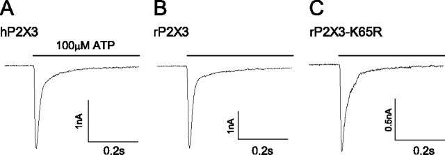
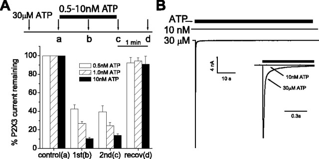
P2X3 receptors desensitize within 100 ms of channel activation, yet recovery from desensitization requires several minutes. The molecular basis for this slow rate of recovery is unknown. We designed experiments to test the hypothesis that this slow recovery is attributable to the high affinity (< 1 nM) of desensitized P2X3 receptors for agonist. We found that agonist binding to the desensitized state provided a mechanism for potent inhibition of P2X3 current. Sustained applications of 0.5 nM ATP inhibited > 50% of current to repetitive applications of P2X3 agonist. Inhibition occurred at 1000-fold lower agonist concentrations than required for channel activation and showed strong use dependence. No inhibition occurred without previous activation and desensitization. Our data are consistent with a model whereby inhibition of P2X3 by nanomolar [agonist] occurs by the rebinding of agonist to desensitized channels before recovery from desensitization. For several ATP analogs, the concentration required to inhibit P2X3 current inversely correlated with the rate of recovery from desensitization. This indicates that the affinity of the desensitized state and recovery rate primarily depend on the rate of agonist unbinding. Consistent with this hypothesis, unbinding of [32P]ATP from desensitized P2X3 receptors mirrored the rate of recovery from desensitization. As expected, disruption of agonist binding by site-directed mutagenesis increased the IC50 for inhibition and increased the rate of recovery.


Figures

Figure 1.
Figure 1.
Representative currents from wild-type and mutant P2X3 receptors. P2X3 receptors from human (A), rat (B), and rat K65R mutation (C) were expressed in HEK cells and currents recorded at -60 mV using whole-cell patch-clamp methods. ATP (100 μm; 0.5 s) application produced a rapidly desensitizing inward current. Mutation of a lysine residue (K65R) slowed the rate of desensitization by twofold (τentry = 26 ± 3 ms; n = 8) when compared with wild-type rat (τentry = 13 ± 1 ms; n = 10) or human (τ = 12 ± 1 ms; n = 8) P2X3 receptors.
Figure 2.
Figure 2.
P2X3 recovery from desensitization depends on agonist structure and is speeded by a binding-site mutation. Saturating (30 μm) ATP (filled circles), αβmeATP (open circles), ATPγS (squares), or CTP (100 μm; triangles) were applied to human (left), rat (middle), or the mutant P2X3-K65R (right) P2X3 receptors to induce desensitization. Current recovery was monitored by reapplication of agonist at the indicated intervals. Note change of time scale for P2X3-K65R curve (right). Data are expressed as a percentage of maximal recovery determined by a fit of the data (see Materials and Methods; n = 4-10 cells for each data point). Error bars represent SEM.
Figure 3.
Figure 3.
The dissociation rate of ATP from the hP2X3 receptor follows the rate of recovery. HEK cells expressing the hP2X3 receptor (filled symbols) or nontranfected HEK cells (open symbols) were incubated with 1 nm [32P]ATP. Unbound [32P]ATP was washed away, and the amount of released [32P]ATP was determined by sampling the bath solution at the times indicated. Released ATP was expressed as a function of total [protein]. The time course of [32P]ATP release was fit with the same equation as recovery from desensitization (see Materials and Methods) and followed a similar time course. Nontransfected cells released little [32P]ATP. Each point represents the mean ± SEM for four independent experiments.
Figure 4.
Figure 4.
Nanomolar [ATP] inhibit the desensitized, but not recovered, P2X3 receptor. A, Saturating [ATP] (30 μm for 0.5 s; arrows) was applied at 1 min intervals. During the 2 min interval between a and c, a low [ATP] (0.5-10 nm) was washed onto the cell. This treatment decreased the current to a similar extent at 1 min (b) and 2 min (c) after the nm ATP wash. Current recovered (recov) completely from inhibition when ATP was removed from the wash during the interval between c and d. B, ATP (solid bar) at 10 nm produced no current when applied for 60 s to a cell expressing hP2X3 (top trace). Immediately after the cessation of 10 nm ATP, the cell was washed for 30 s with an ATP-free solution. ATP (30 μm) applied after washout produced a large inward current (bottom trace), indicating that the cell was responsive to higher [ATP]. The two currents were superimposed on a shorter time scale (inset). Data are expressed as mean peak currents ± SEM (n = 10-12).
Figure 5.
Figure 5.
Potency of P2X3 inhibition diminishes as channels recover from desensitization. A, 100 μm ATPγS (arrows; first application not shown) was applied for 0.5 s at 60 s intervals to HEK cells that expressed the hP2X3 receptor. During the first or second half of the interval between applications b and c, 1-100 nm ATP was applied for 30 s. Inhibition was monitored as the fraction of current induced after the test ATP treatment (current at c/current at b). B shows a representative experiment in which 10 nm ATP was applied “early” in the interval. The current to subsequent 100 μm ATPγS (c) was diminished. (Current time scale is expanded from A to visualize P2X3 current; axis break, 59 s.) C, Summary of experiments as in B. Application of ATP during the first 30 s of the interval (early; triangles) was more effective at inhibiting P2X3 current than when applied for 30 s immediately before the test ATPγS application (“late”; squares). Data are expressed as average peak current ratios ± SEM (n = 6-11 cells for each data point).
Figure 6.
Figure 6.
Kinetics of onset and offset of inhibition by nanomolar agonist. A, CTP (100 μm) was applied for 0.5 s at 15 s intervals to activate and desensitize hP2X3 current. This protocol results in a stable inward current (first 3 deflections). Application of 3 nm ATP (120 s) decreased current to CTP > 95% within 100 s (n = 12). After washout of ATP, the current to CTP recovered. Onset of inhibition was faster than offset. B, Inhibition by nanomolar ATP was use dependent. Application of 3 nm ATP (120 s) did not inhibit current when applied without CTP-induced channel activation and subsequent desensitization (n = 8). C, The ATP analog αβmeATP (3 nm for 120 s) was less effective and had a slower onset than ATP at inhibiting current (n = 11). Current recovery from αβmeATP-induced inhibition was faster than with ATP.
Figure 7.
Figure 7.
Agonist that promote faster recovery are less potent inhibitors of P2X3 current. A, Half-maximal inhibition occurs at >400-fold lower concentrations than activation. For inhibition curves (filled symbols), 100 μm ATPγS was applied at 60 s intervals to evoke P2X3 current. After a stable current is established, the indicated concentration of nucleotide, ATP (•), αβmeATP (▴), ATPγS(▪), or CTP (▾), was applied for 60 s. The decrease in 100 μm ATPγS-induced current immediately after the treatment interval is plotted against agonist concentration and fitted with the Hill equation (curves). Dose-response for activation of P2X3 current is plotted on the same graph (open symbols). Dose-response data were collected at 60 s intervals for all agonists except ATP (120 s interval). Experiment was repeated using the binding site mutant P2X3-K65R and ATP as desensitizing (•) or activating (○) agonist (dashed lines). B, IC50 values for inhibition of current positively correlate to the rate of recovery from desensitization. Like low-potency agonists, the K65R mutation increases rate of recovery and decreases ability to promote desensitization. Error bars represent SEM.

References

    1. Alexander K, Niforatos W, Bianchi B, Burgard EC, Lynch KJ, Kowaluk EA, Jarvis MF, van Biesen T (1999) Allosteric modulation and accelerated resensitization of human P2X(3) receptors by cibacron blue. J Pharmacol Exp Ther 291: 1135-1142. - PubMed
    1. Bartrup JT, Newberry NR (1996) Electrophysiological consequences of ligand binding to the desensitized 5-HT3 receptor in mammalian NG108-15 cells. J Physiol (Lond) 490: 679-690. - PMC - PubMed
    1. Chang Y, Ghansah E, Chen Y, Ye J, Weiss DS (2002) Desensitization mechanism of GABA receptors revealed by single oocyte binding and receptor function. J Neurosci 22: 7982-7990. - PMC - PubMed
    1. Changeux JP (1990) The TiPS lecture. The nicotinic acetylcholine receptor: an allosteric protein prototype of ligand-gated ion channels. Trends Pharmacol Sci 11: 485-492. - PubMed
    1. Chen CC, Akopian AN, Sivilotti L, Colquhoun D, Burnstock G, Wood JN (1995) A P2X purinoceptor expressed by a subset of sensory neurons. Nature 377: 428-431. - PubMed

Substances