. 2005 Aug 10;25(32):7366-76.
doi: 10.1523/JNEUROSCI.0899-05.2005.

Coupled networks of parvalbumin-immunoreactive interneurons in the rat basolateral amygdala

Affiliations

Coupled networks of parvalbumin-immunoreactive interneurons in the rat basolateral amygdala

Jay F Muller et al. J Neurosci. .

Abstract

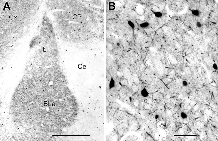
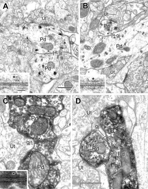
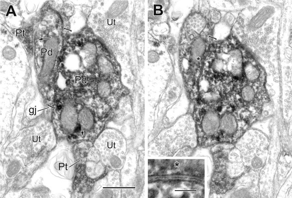
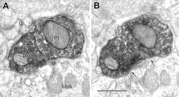
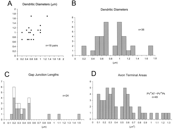
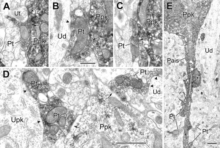
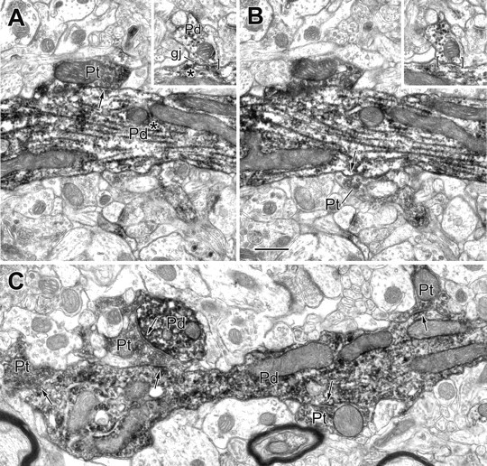
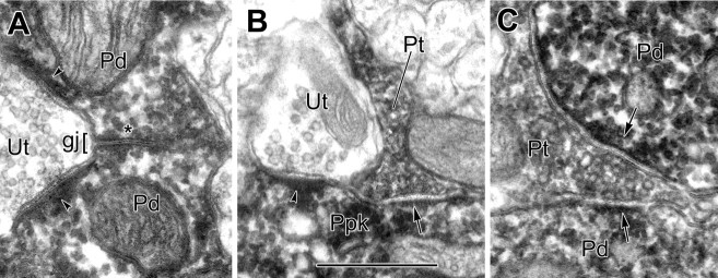
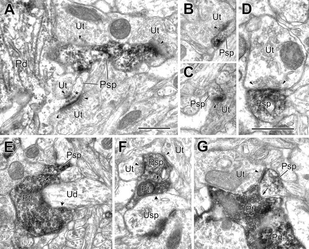
Recent studies indicate that the basolateral amygdala exhibits fast rhythmic oscillations during emotional arousal, but the neuronal mechanisms underlying this activity are not known. Similar oscillations in the cerebral cortex are generated by a network of parvalbumin (PV)-immunoreactive interneurons interconnected by chemical synapses and dendritic gap junctions. The present immunoelectron microscopic study revealed that the basolateral amygdalar nucleus (BLa) contains a network of parvalbumin-immunoreactive (PV+) interneurons interconnected by chemical synapses, dendritic gap junctions, and axonal gap junctions. Twenty percent of synapses onto PV+ neurons were formed by PV+ axon terminals. All of these PV+ synapses were symmetrical. PV+ perikarya exhibited the greatest incidence of PV+ synapses (30%), with lower percentages associated with PV+ dendrites (15%) and spines (25%). These synapses comprised half of all symmetrical synapses formed with PV+ cells. A total of 18 dendrodendritic gap junctions between PV+ neurons were observed, mostly involving secondary and more distal dendrites (0.5-1.0 microm thick). Dendritic gap junctions were often in close proximity to PV+ chemical synapses. Six gap junctions were observed between PV+ axon terminals. In most cases, one or both of these terminals formed synapses with the perikarya of principal neurons. This is the first study to describe dendritic gap junctions interconnecting PV+ interneurons in the basolateral amygdala. It also provides the first documentation of gap junctions between interneuronal axon terminals in the mammalian forebrain. These data provide the anatomical basis for a PV+ network that may play a role in the generation of rhythmic oscillations in the BLa during emotional arousal.


Figures

Figure 1.
Figure 1.
Light micrographs of parvalbumin immunoreactivity. A, A low-magnification micrograph of a coronal section through the rat amygdala (bregma level, -2.7) showing PV immunoreactivity in the BLa and adjacent regions (Ce, central amygdalar nucleus; L, lateral amygdalar nucleus; Cx, cortex; CP, caudatoputamen). B, A higher-magnification view showing ∼90% of an area remounted and thin sectioned for quantitative electron microscopic analysis (see Materials and Methods). Scale bars: A, 500 μm; B, 50 μm.
Figure 2.
Figure 2.
Electron micrographs of gap junctions between PV+ dendrites in the BLa. A, A gap junction (gj; bracket), 0.22 μm in length, between two PV+ dendrites (Pd), labeled using the pre-embedding IGS technique. One labeled dendrite receives synaptic input from an unlabeled terminal (Ut). The asterisk indicates the site of gap junctional contact, which is enlarged in the inset. B, A gap junction (gj; bracket), 0.2 μm in length, between two PV+ dendrites (Pd), is labeled using the V-VIP substrate technique for peroxidase. The asterisk indicates the area of contact, which is enlarged in the inset. Both labeled dendrites received synaptic input from unlabeled terminals (Ut). In serial sections, one terminal (top, left) was seen to form symmetrical contacts with both dendrites, whereas the other terminals formed asymmetrical synapses. C, A gap junction (gj; bracket), 0.33 μm in length, between two PV+ dendrites (Pd), is labeled using the immunoperoxidase technique. The asterisk indicates the area of contact, which is enlarged in the inset. An unlabeled terminal (Ut) makes asymmetrical synaptic contact onto both labeled dendrites (arrowheads). D, A gap junction (gj; brackets), 0.5 μm in length, between two PV+ dendrites (Pd), is labeled using the IP technique. Both labeled dendrites receive synaptic input from unlabeled terminals (Ut; arrowheads). Scale bars: 0.5 μm; insets, 50 nm. B is the same magnification as A. C is the same magnification as D.
Figure 3.
Figure 3.
Two serial electron micrographs of a gap junction (gj; brackets) between two PV+ dendrites (Pd), both receiving synaptic input from PV+ terminals (Pt; arrows). The gap junction, followed for five serial sections, measured 0.9 μm in length. The asterisk in B marks the gap junction region, which is enlarged in the inset. Unlabeled terminals (Ut) are also in contact with the larger labeled dendrite. Scale bars: A, B, 0.5 μm; inset, 100 nm.
Figure 4.
Figure 4.
A, B, Two serial electron micrographs of a gap junction (gj; brackets) between two PV+ axon terminals (Pt). The gap junction, followed through four serial sections, measured 0.46 μm in length. Both labeled terminals synapse onto the same PV-negative perikaryon of a pyramidal cell (arrows; Upk). Scale bar, 0.5 μm.
Figure 5.
Figure 5.
Graphic representation of measured profiles. A, A scatter plot of the diameters of the 18 pairs of PV+ dendrites in the BLa forming gap junctions, measured as the largest diameter in the vicinity of the gap junction. The y-axis represents the larger-caliber dendrite, where the diameters differ. B, A histogram representing diameters of the individual dendrites plotted as pairs in A. C, A histogram of the measured lengths of gap junctions. Gray bars represent the gap junctions between PV+ dendrites plotted in A and B (above; n = 18). The white bars add the lengths of the six gap junctions observed between PV+ axon terminals (n = 6). All junctions but one were followed, at least partially, through serial sections. D, Histogram of the areas of 49 PV+ terminals found to synapse onto the 16 PV+ perikarya of the quantitative survey.
Figure 6.
Figure 6.
Electron micrographs of synapses from PV+ terminals onto PV+ perikarya and an axon initial segment. A, A PV+ perikaryon (Ppk) receives synaptic input from a PV+ terminal (Pt; arrow) and asymmetrical synaptic contact from an adjacent unlabeled terminal (Ut; arrowhead). B, C, Examples of two PV+ terminals (Pt; arrows) synapsing onto the same PV+ perikaryon (Ppk). In B, the larger PV+ terminal also makes synaptic contact onto an unlabeled proximal dendrite of a pyramidal cell (Ud; short arrow). D, Two PV+ terminals make synaptic contact onto the same moderately labeled PV+ perikaryon (Ppk) that was originally identified at the light microscopic level (long arrows). The larger PV+ terminal at the left is also one of two adjacent PV+ terminals synapsing onto an unlabeled perikaryon of a pyramidal (Upk; short arrows). At the right, the smaller PV+ terminal is one of two PV+ terminals making synaptic contact with the same unlabeled dendrite (Ud; short arrows). E, Axon initial segment (Pais) arising from a PV+ perikaryon receives synaptic contact from four small (Pt) and one large PV+ terminal (asterisk). The latter also makes synaptic contact with a large-caliber PV-negative dendrite (Ud). Scale bars: (in B) A-C, 0.5 μm; D, E, 1 μm.
Figure 7.
Figure 7.
Electron micrographs of PV+ dendrites exhibiting a gap junction and receiving synapses from PV+ terminals. A, B, Two serial sections of a large-caliber PV+ dendrite (Pd) showing two of three PV+ terminals (Pt; arrows) that make synaptic contact with this dendrite. The inset depicts a gap junction (gj) involving this dendrite and a small PV+ dendrite (Pd; brackets) that was located 11 μm from the synapses. The asterisks indicate that all contacts are with the same PV+ dendritic trunk. C, Longitudinal view of a varicose PV+ dendrite (Pd) receiving synaptic input from four PV+ terminals (Pt; arrows). One of these PV+ terminals also synapses onto an adjacent PV+ dendritic cross section (Pd; arrow; top left). Scale bar, 0.5 μm.
Figure 8.
Figure 8.
Comparison of the morphology of an IP-labeled gap junction with IP-labeled chemical synapses. A, Detail of the IP-labeled PV+ dendrites (Pd) shown in Figure 2C in gap junctional contact (gj; asterisk). Each dendrite receives an asymmetrical synapse (arrowheads) from an unlabeled terminal (Ut). B, Detail from Figure 6 A of an IP-labeled PV+ perikaryon (Ppk) receiving synaptic contacts from a PV+ axon terminal (Pt; arrow) and unlabeled axon terminal (Ut; arrowhead). C, Detail from Figure 7C of an IP-labeled PV+ terminal (Pt) making synaptic contacts (arrows) with two PV+ dendrites (Pd). Note that peroxidase reaction product fills the central gap of the gap junction, whereas an unstained synaptic cleft separates the outer leaflets of presynaptic and postsynaptic plasma membranes of chemical synapses. Scale bar: (in B) A-C, 0.5 μm.
Figure 9.
Figure 9.
Electron micrographs of PV+ spines receiving synaptic input from both PV-negative and PV+ terminals. A, Two spines (Psp) emerge from a PV+ dendrite (Pd), each receiving synaptic input from two unlabeled terminals (Ut; arrowheads). In serial sections, all contacts appeared asymmetrical. One of the spines has subsynaptic dense bodies (asterisk). B, C, Two neighboring PV+ spines (Psp) receive synaptic input from unlabeled terminals (Ut; arrowheads). D, A higher-magnification view of a PV+ spine (Psp) receiving unlabeled synaptic input (Ut; arrowheads). E, A PV+ spine (Psp) receives synaptic input from a PV+ terminal (Pt; long arrow) that is also wrapped around and synapsing onto an unlabeled dendrite (Ud; short arrow). F, A PV+ spine (Psp) receives synaptic input from a PV+ terminal (Pt; long arrow) and two unlabeled terminals (Ut; arrowheads). The PV+ terminal also makes synaptic contact onto an unlabeled spine (Usp; short arrow). G, APV+ spine (Psp) receives synaptic input from PV+ (Pt; arrow) and unlabeled (Ut; arrowhead) terminals. The PV+ terminal appears to be contacting a PV+ dendrite (Pd). A-C and E-G are the same magnification. Scale bars, 0.5 μm.

Similar articles

Cited by

References

    1. Aggleton JP (1992) The amygdala: neurobiological aspects of emotion, memory, and mental dysfunction. New York: Wiley.
    1. Aggleton JP (2000) The amygdala: a functional analysis. New York: Wiley.
    1. Baldridge WH, Ball AK, Miller RG (1987) Dopaminergic regulation of horizontal cell gap junction particle density in goldfish retina. J Comp Neurol 265: 428-436. - PubMed
    1. Bartos M, Vida I, Frotscher M, Meyer A, Monyer H, Geiger JR, Jonas P (2002) Fast synaptic inhibition promotes synchronized gamma oscillations in hippocampal interneuron networks. Proc Natl Acad Sci USA 99: 13222-13227. - PMC - PubMed
    1. Belluardo N, Mudò G, Trovato-Salinaro A, Gurun SL, Charollais A, Serre-Beinier V, Amato G, Haefliger J-A, Meda P, Condorelli DF (2000) Expression of connexin 36 in the adult and developing rat brain. Brain Res 865: 121-138. - PubMed

Publication types

MeSH terms

LinkOut - more resources