

How to enhance ipsilateral actions of pyramidal tract neurons
- PMID: 16093391
- PMCID: PMC1890015
- DOI: 10.1523/JNEUROSCI.1838-05.2005
How to enhance ipsilateral actions of pyramidal tract neurons
Abstract
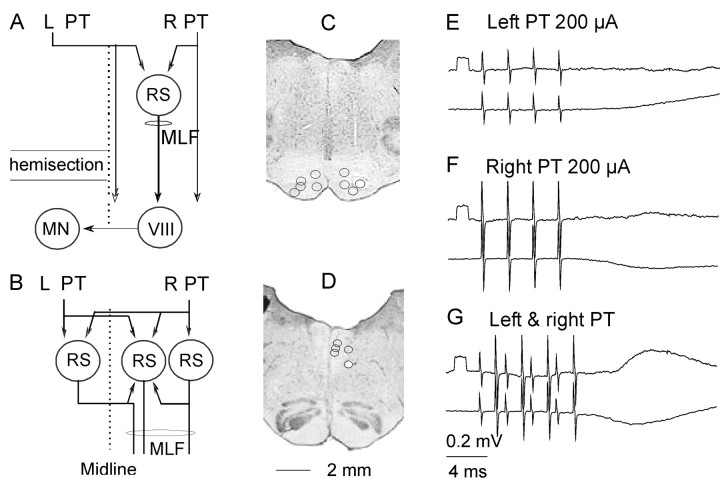
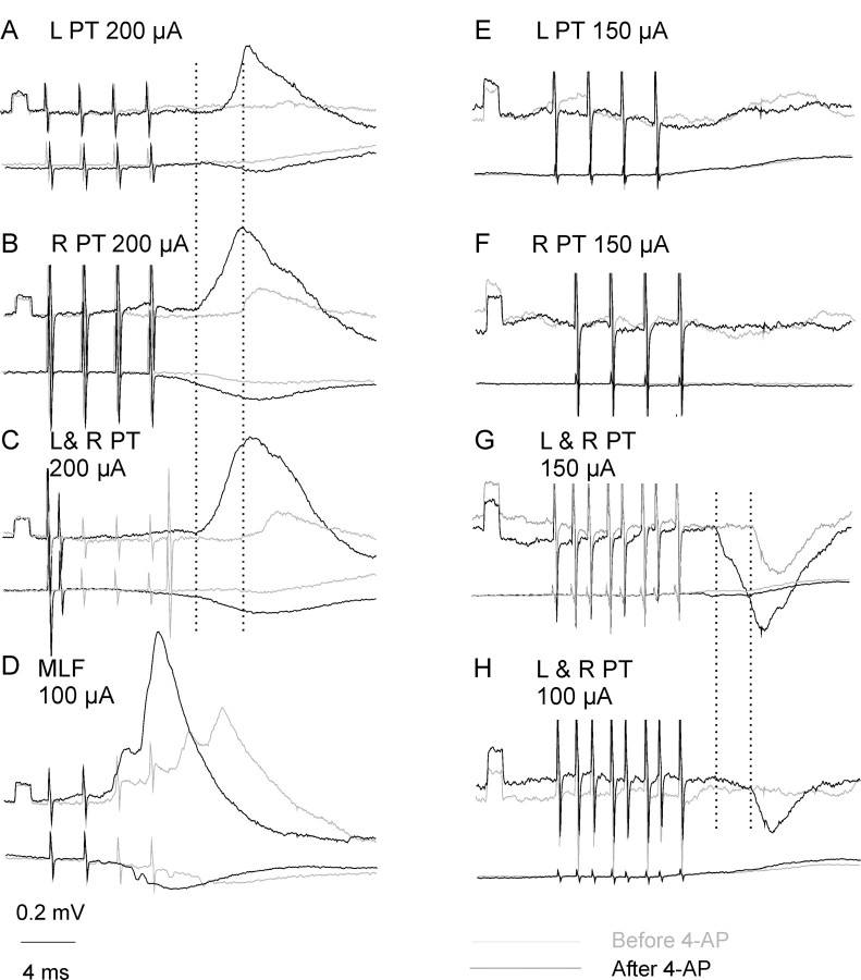
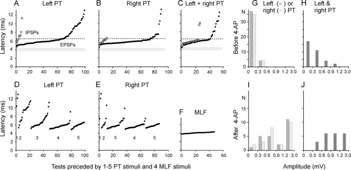
We have shown previously that ipsilateral pyramidal tract (PT) neurons facilitate the actions of reticulospinal neurons on feline motoneurons (Edgley et al., 2004), which indicates that they might assist the recovery of motor functions after injuries of contralateral corticospinal neurons. Nevertheless, stimulation of ipsilateral PT fibers alone only rarely evoked any synaptic actions in motoneurons. The aim of this study was to investigate possible ways of enhancing such actions and of inducing more effective excitation and inhibition of motoneurons. The effects of stimulation of the ipsilateral PT were investigated after eliminating the spinal actions of contralateral PT fibers by hemisecting the spinal cord at a low thoracic level and were estimated from intracellular records from hindlimb motoneurons. Two measures were used to enhance PT actions. The first was to increase the probability of activation of reticulospinal neurons by mutual facilitation of actions of ipsilateral and contralateral PT neurons. The second was to enhance synaptic transmission between PT neurons and reticulospinal neurons, and in pathways between the reticulospinal neurons and motoneurons via commissural interneurons, by systemic application of a K+ channel blocker, 4-aminopyridine (4-AP). The results show that under favorable conditions, ipsilateral PT neurons may induce EPSPs and IPSPs in hindlimb motoneurons, or even action potentials, via the reticulospinal pathway. This study strengthens previous conclusions that ipsilateral PT neurons can potentially replace, at least to some extent, the actions of injured contralateral PT neurons. It also suggests that 4-AP might improve the progress of the recovery.
Figures

Similar articles
-
Ipsilateral actions of feline corticospinal tract neurons on limb motoneurons.J Neurosci. 2004 Sep 8;24(36):7804-13. doi: 10.1523/JNEUROSCI.1941-04.2004. J Neurosci. 2004. PMID: 15356191 Free PMC article.
-
Ipsilateral actions from the feline red nucleus on hindlimb motoneurones.J Physiol. 2008 Dec 15;586(24):5865-84. doi: 10.1113/jphysiol.2008.163998. Epub 2008 Oct 20. J Physiol. 2008. PMID: 18936076 Free PMC article.
-
Are crossed actions of reticulospinal and vestibulospinal neurons on feline motoneurons mediated by the same or separate commissural neurons?J Neurosci. 2003 Sep 3;23(22):8041-50. doi: 10.1523/JNEUROSCI.23-22-08041.2003. J Neurosci. 2003. PMID: 12954866 Free PMC article.
-
[Functional organization of segmentally homologous reticulospinal neurons in hindbrain].Tanpakushitsu Kakusan Koso. 2004 Feb;49(3 Suppl):486-92. Tanpakushitsu Kakusan Koso. 2004. PMID: 14976777 Review. Japanese. No abstract available.
-
Physiological basis of motor effects of a transient stimulus to cerebral cortex.Neurosurgery. 1987 Jan;20(1):74-93. Neurosurgery. 1987. PMID: 3543727 Review.
Cited by
-
Neuronal relays in double crossed pathways between feline motor cortex and ipsilateral hindlimb motoneurones.J Physiol. 2006 Sep 1;575(Pt 2):527-41. doi: 10.1113/jphysiol.2006.112425. Epub 2006 Jun 1. J Physiol. 2006. PMID: 16740611 Free PMC article.
-
Distribution of 28 kDa Calbindin-Immunopositive Neurons in the Cat Spinal Cord.Front Neuroanat. 2016 Jan 28;9:166. doi: 10.3389/fnana.2015.00166. eCollection 2015. Front Neuroanat. 2016. PMID: 26858610 Free PMC article.
-
Lack of evidence for direct corticospinal contributions to control of the ipsilateral forelimb in monkey.J Neurosci. 2011 Aug 3;31(31):11208-19. doi: 10.1523/JNEUROSCI.0257-11.2011. J Neurosci. 2011. PMID: 21813682 Free PMC article.
-
Muscle agonist-antagonist interactions in an experimental joint model.Exp Brain Res. 2012 Oct;222(4):399-414. doi: 10.1007/s00221-012-3227-0. Epub 2012 Aug 29. Exp Brain Res. 2012. PMID: 22926155 Free PMC article.
-
Functional plasticity of the ipsilateral primary sensorimotor cortex in an elite long jumper with below-knee amputation.Neuroimage Clin. 2019;23:101847. doi: 10.1016/j.nicl.2019.101847. Epub 2019 May 9. Neuroimage Clin. 2019. PMID: 31103873 Free PMC article.
References
-
- Bawa P, Hamm JD, Dhillon P, Gross PA (2004) Bilateral responses of upper limb muscles to transcranial magnetic stimulation in human subjects. Exp Brain Res 158: 385-390. - PubMed
-
- Caramia MD, Iani C, Bernardi G (1996) Cerebral plasticity after stroke as revealed by ipsilateral responses to magnetic stimulation. NeuroReport 7: 1756-1760. - PubMed
-
- Hallett M (2001) Functional reorganization after lesions of the human brain: studies with transcranial magnetic stimulation. Rev Neurol (Paris) 157: 822-826. - PubMed
Publication types
MeSH terms
Substances
Grants and funding
LinkOut - more resources
Full Text Sources