. 2005 Aug 10;25(32):7406-19.
doi: 10.1523/JNEUROSCI.1008-05.2005.

Mechanisms of neuropeptide Y, peptide YY, and pancreatic polypeptide inhibition of identified green fluorescent protein-expressing GABA neurons in the hypothalamic neuroendocrine arcuate nucleus

Affiliations

Mechanisms of neuropeptide Y, peptide YY, and pancreatic polypeptide inhibition of identified green fluorescent protein-expressing GABA neurons in the hypothalamic neuroendocrine arcuate nucleus

Claudio Acuna-Goycolea et al. J Neurosci. .

Abstract

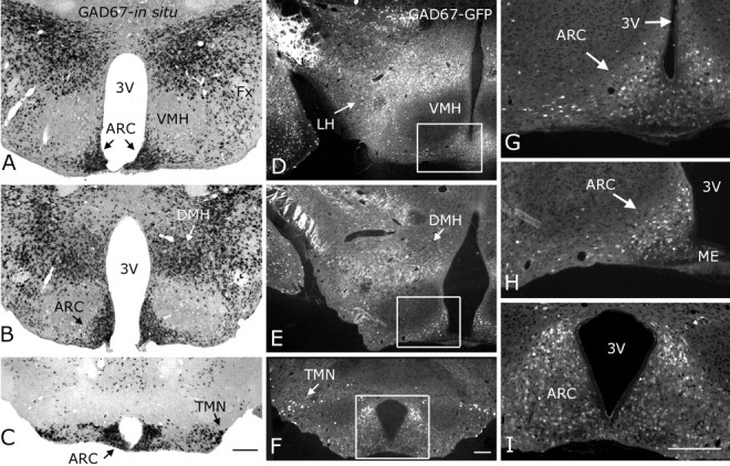
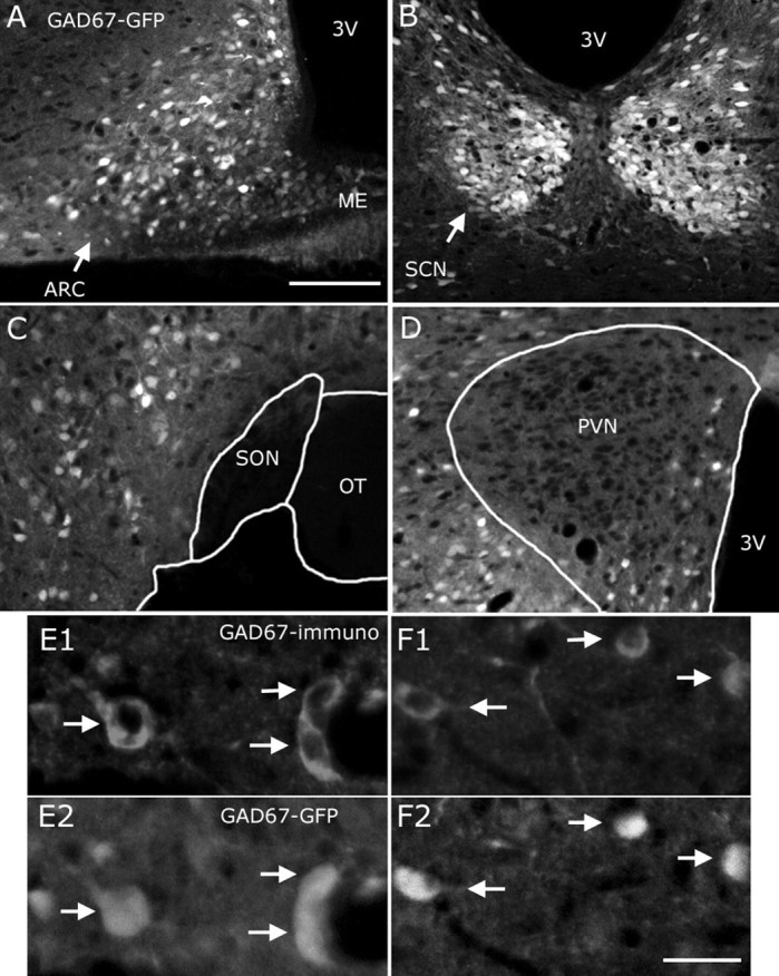
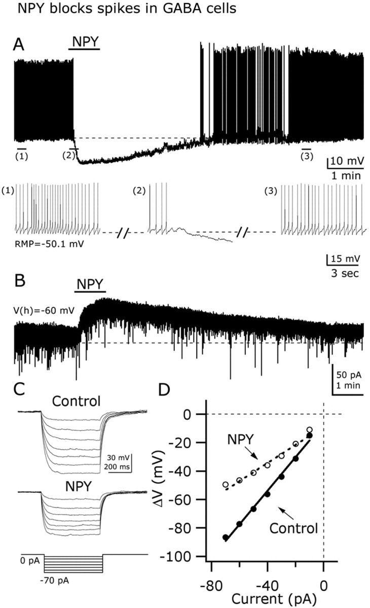
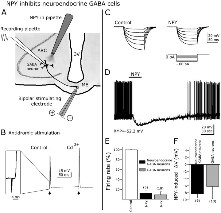
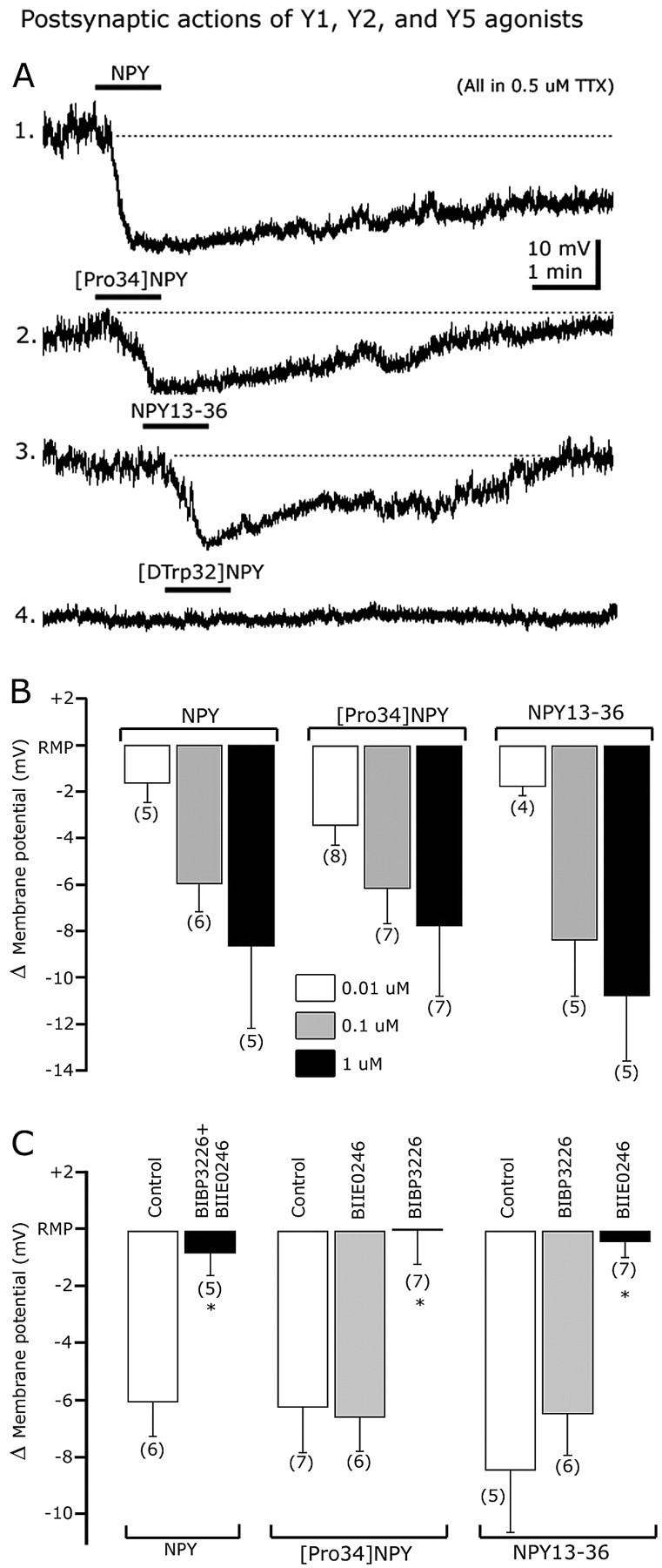
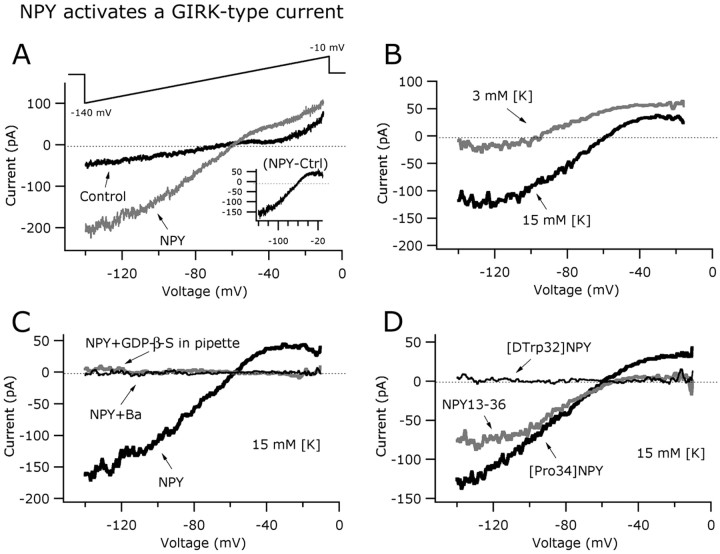
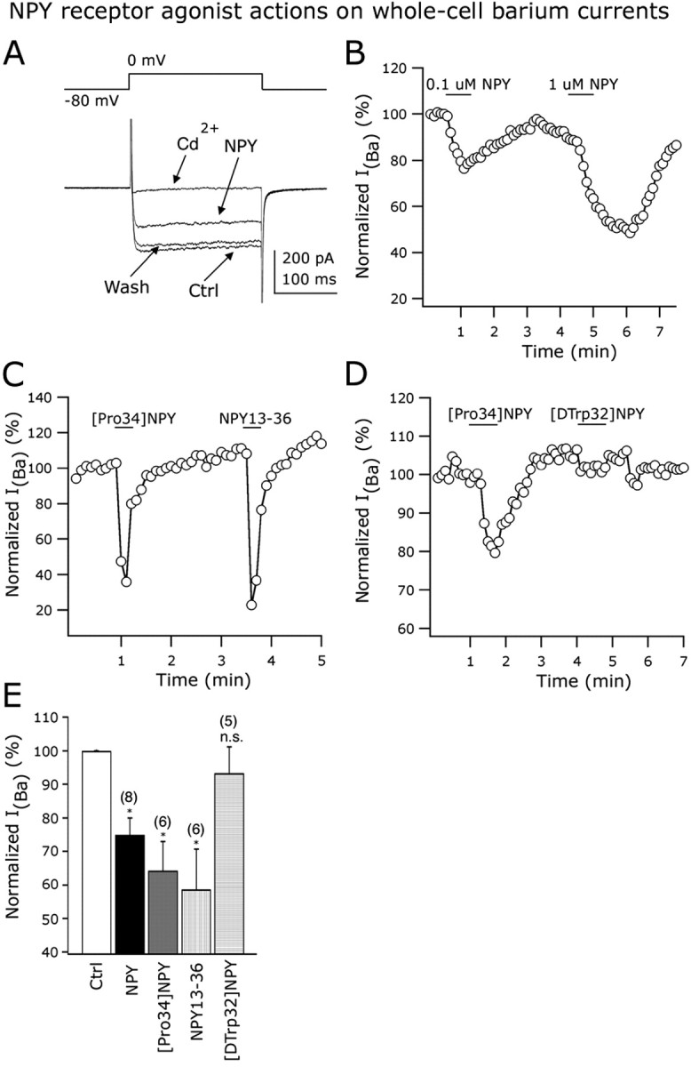
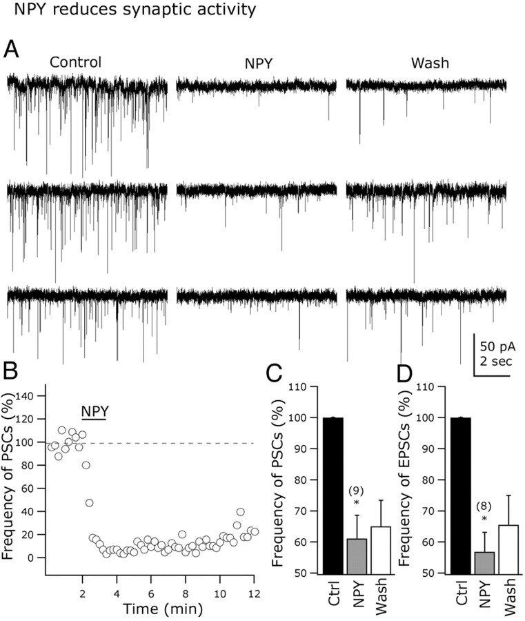
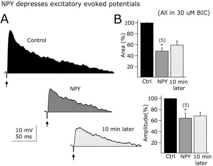
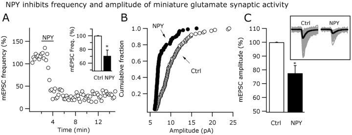
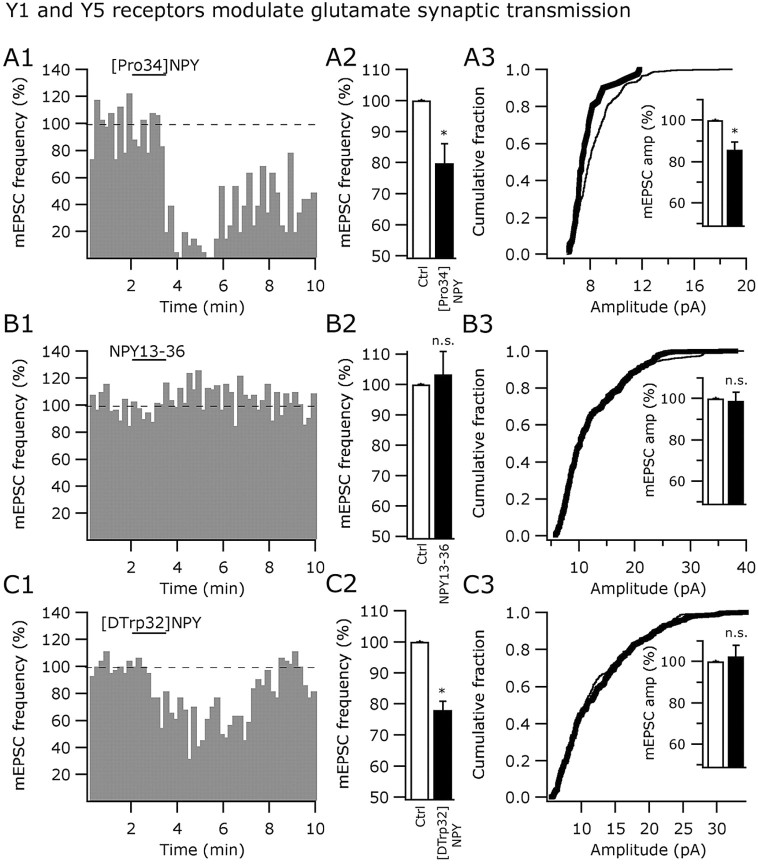
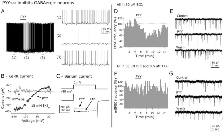
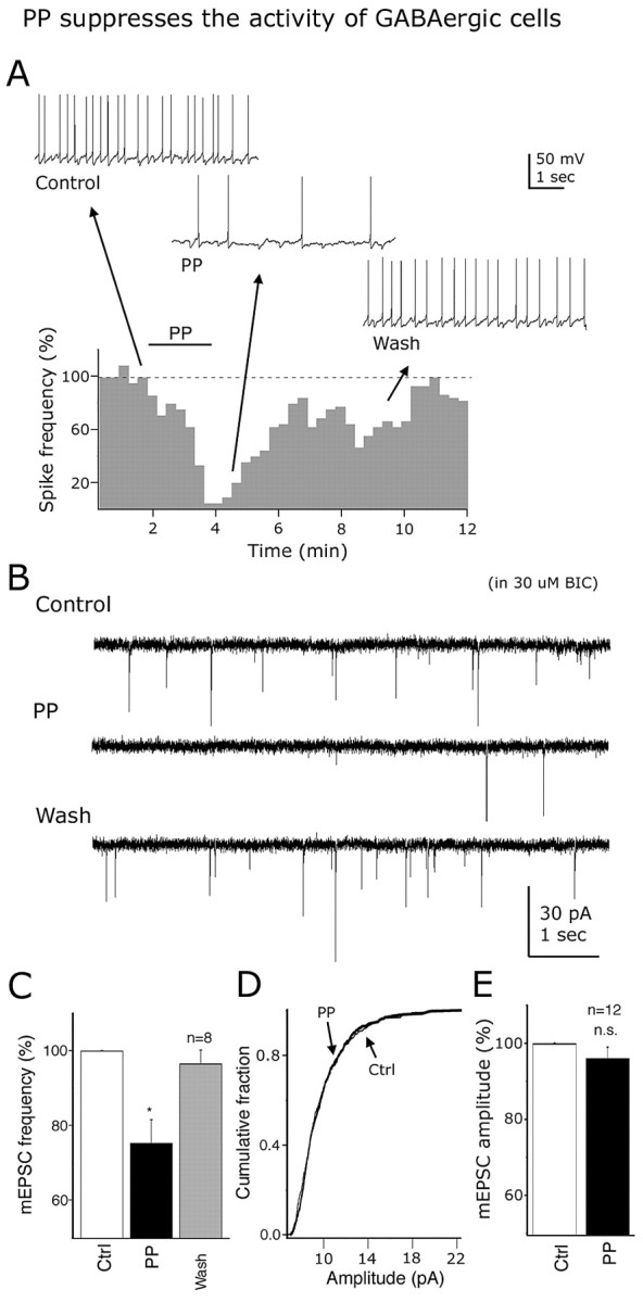
The fast inhibitory transmitter GABA is robustly expressed in the arcuate nucleus (ARC) and appears to play a major role in hypothalamic regulation of endocrine function and energy homeostasis. Previously, it has not been possible to record selectively from GABA cells, because they have no defining morphological or physiological characteristics. Using transgenic mice that selectively express GFP (green fluorescent protein) in GAD67 (glutamic acid decarboxylase 67)-synthesizing cells, we identified ARC GABA neurons (n > 300) and used whole-cell recording to study their physiological response to neuropeptide Y (NPY), the related peptide YY(3-36) (PYY(3-36)), and pancreatic polypeptide (PP), important modulators of ARC function. In contrast to other identified ARC cells in which NPY receptor agonists were reported to generate excitatory actions, we found that NPY consistently reduced the firing rate and hyperpolarized GABA neurons including neuroendocrine GABA neurons identified by antidromic median eminence stimulation. The inhibitory NPY actions were mediated by postsynaptic activation of G-protein-linked inwardly rectifying potassium (GIRK) and depression of voltage-gated calcium currents via Y1 and Y2 receptor subtypes. Additionally, NPY reduced spontaneous and evoked synaptic glutamate release onto GABA neurons by activation of Y1 and Y5 receptors. The peptide PYY(3-36), a peripheral endocrine signal that can act in the brain, also inhibited GABA neurons, including identified neuroendocrine cells, by activating GIRK conductances and depressing calcium currents. The endogenous Y4 agonist PP depressed the activity of GABA-expressing neurons mainly by presynaptic attenuation of glutamate release. Together, these results show that the family of neuropeptide Y modulators reduces the activity of inhibitory GABA neurons in the ARC by multiple presynaptic and postsynaptic mechanisms.


Figures

Figure 1.
Figure 1.
GAD67 mRNA matches GAD67-GFP in transgenic mice. A-C, In situ hybridization showing GAD67 mRNA (small black spots) was strongly expressed in ARC, lateral hypothalamus, dorsomedial nucleus, and tuberomammillary nucleus in the posterior hypothalamus. Very low levels of GAD67 mRNA were found in the ventromedial hypothalamic nucleus. D-F, GFP-producing cells (GFP shown in white) were detected in the same hypothalamic regions where GAD67 mRNA was found. G-I, High-power magnification of the ARC boxed areas in D-E, respectively. Scale bars: C (for A-C), F (for D-F), I (for G-I), 200 μm. 3V, Third ventricle; Fx, fornix.
Figure 2.
Figure 2.
Colocalization of GFP and GAD67 immunoreactivity in the hypothalamus of GAD67-GFP mice. In addition to the ARC (A), GFP was also detected in the suprachiasmatic nucleus (B) in the transgenic mice. GFP was found in neither the supraoptic (C) nor the magnocellular area of the paraventricular hypothalamic nucleus (D). In both the medial (E1-E2, white arrows) and lateral (F1-F2, white arrows) hypothalamus, GFP colocalized with GAD67 immunoreactivity, consistent with the selective GFP expression in GAD67-producing neurons. In this figure, both green (GFP expression) and red (Texas Red from secondary antibody) fluorescence are presented in white. Scale bars: (in A) A-D, 200 μm; (in F2) E1-F2, 20 μm. 3V, Third ventricle; OT, optic tract.
Figure 3.
Figure 3.
NPY inhibits GAD67-expressing neurons in arcuate nucleus. A, Current-clamp recording from a representative neuron showing the inhibitory effect of 1 μm NPY on the frequency of action potentials. A hyperpolarizing shift in the membrane potential is also evident after NPY application. Extended traces before, during, and after NPY are depicted in the bottom part (1, 2, and 3 indicate selected parts of the trace). B, In voltage-clamp (Vh = -60 mV), NPY evoked a robust and long-lasting outward current. C, NPY decreased the voltage response of GAD67 neurons after hyperpolarizing current steps (left) (see protocol in the bottom part). D, This graph shows the current-voltage relationship in the absence (right, filled circles) and the presence (white circles) of NPY. NPY evoked a decrease in the whole-cell input resistance (slope). RMP, Resting membrane potential.
Figure 4.
Figure 4.
NPY inhibits ARC neuroendocrine GABAergic neurons. A, Schematic representation of hypothalamic slice preparation used to study neuroendocrine cells. 3V, Third ventricle. B, Antidromic responses (action potentials) were evoked in GFP-expressing cells after median eminence electrical stimulation with a bipolar electrode. The action potentials induced by ME antidromic stimulation persisted in 200 μm CdCl2 in the bath, indicating that these responses were not attributable to transmitter release induced by orthodromic activation of recurrent axon collaterals. Inset shows the spike latency and initial rise of an action potential evoked by ME stimulation. C, In this typical cell, negative current steps (see protocol in the bottom part) were delivered before (control) and during NPY application. A robust reduction in the hyperpolarizing responses after negative current injection was observed in the presence of NPY, consistent with a decrease in whole-cell input resistance. D, In current-clamp conditions, NPY blocked spikes and hyperpolarized ARC neuroendocrine neurons. E, F, NPY-induced reduction in spike frequency and hyperpolarization in neuroendocrine neurons were not significantly different from that of other ARC GABA cells. Error bars indicate SE. The number of cells is shown in parentheses.
Figure 5.
Figure 5.
Y1 and Y2, but not Y5, receptor subtypes are involved in the direct dose-dependent postsynaptic actions of NPY. A, Representative traces showing the inhibitory effect of NPY (trace 1), [Pro34]NPY (a selective Y1 agonist) (trace 2), and NPY13-36 (a Y2 agonist) (trace 3) on the membrane potential of identified GAD67 neurons. The Y5 receptor agonist [d-Trp32]NPY1-36 (trace 4) did not alter the membrane potential. B, Dose-dependent effects of NPY (left), [Pro34]NPY1-36 (middle), and NPY13-36 (right) on membrane potential. C, Bar graph summarizing Y1 and Y2 agonist and antagonist actions on hyperpolarization induced by NPY (left), [Pro34]NPY1-36 (middle), and NPY13-36 (right). Error bars indicate SE. *Statistically significant. The number of cells is shown in parentheses.
Figure 6.
Figure 6.
NPY activates a GIRK-like current in arcuate GABA neurons. A, Typical current responses of an identified GAD67 cell before (black trace) and during (gray trace) the application of NPY (1 μm) to the slice. The voltage-ramp protocol used in this experiment is shown in the top part. The net NPY-induced current (inset) was obtained by subtracting control from NPY components. B, NPY-induced current was altered by changing extracellular K+ from 3 to 15 mm. A positive shift in the reversal potential was evident for 15 mm potassium in the bath. C, The current evoked by NPY was G-protein activation dependent, and sensitive to 800 μm barium in the external solution, consistent with a GIRK-type conductance. D, Similarly, both the selective Y1 and Y2 receptor agonists evoked inwardly rectifying currents in arcuate GAD67 neurons. A Y5 receptor agonist, [d-Trp32]NPY1-36, had no effect on the GIRK-like current in these hypothalamic neurons.
Figure 7.
Figure 7.
Y1 and Y2 receptors inhibit voltage-dependent calcium channels. External calcium was replaced by barium in all of these experiments. A, A representative neuron showing the inhibitory effect of 1 μm NPY on the whole-cell IBa. Bath application of 200 μm CdCl2 abolished the IBa, consistent with the idea that IBa was mediated by voltage-dependent calcium channels. B, Inhibitory actions of 0.1 and 1 μm NPY on the normalized amplitude of IBa. C, The Y1 ([Pro34]NPY) and Y2 (NPY13-36) agonists inhibited whole-cell IBa in a single GAD67 neuron from the ARC. D, The Y5 receptor agonist [d-Trp32]NPY1-36 did not evoke a reduction in the IBa amplitude. In the same cell, [Pro34]NPY inhibited IBa. E, Bar graph summarizing the NPY receptor agonist actions on whole-cell IBa in identified arcuate GAD67-containing neurons. Error bars indicate SE. Ctrl, Control; n.s., nonsignificant. *Statistically significant. The number of cells is shown in parentheses.
Figure 8.
Figure 8.
NPY modulates spontaneous excitatory transmission in arcuate nucleus. A, Representative cells showing the NPY-evoked reduction in postsynaptic current frequency. B, This graph shows the time course of the inhibitory NPY effect on the frequency of postsynaptic currents (PSCs) in a representative GAD67 neuron from the ARC. C, D, Bar graphs presenting the mean reduction in the PSC (C) and EPSC (D) frequency with NPY application. Error bars indicate SE. Ctrl, Control. *Statistically significant. The number of cells is shown in parentheses.
Figure 9.
Figure 9.
Excitatory evoked potentials are depressed by NPY. A, Representative evoked potentials before (top), during (middle), and after (bottom) NPY application in a typical GFP-expressing neuron from the ARC. B, Bar graphs show the suppressing NPY effects on the time-voltage integral (top) and peak amplitude (bottom) of the electrically evoked responses. Error bars indicate SE. Ctrl, Control. *Statistically significant. The number of cells is shown in parentheses.
Figure 10.
Figure 10.
NPY actions on miniature glutamatergic activity. A, NPY (1 μm) reduced the frequency of mEPSCs as shown in this time course graph. The inset bar graph shows the mean NPY effect on mEPSC frequency. B, Cumulative distributions of mEPSC amplitude were left shifted in NPY. C, The mean amplitude of the mEPSCs was also reduced by this peptide. The inset shows miniature events in control and NPY conditions. Thicker black traces show the averaged events. Error bars indicate SE. Ctrl, Control. *Statistically significant.
Figure 11.
Figure 11.
Y5 and Y1 receptors modulate glutamate transmission by presynaptic mechanisms. A1-A3, Inhibitory actions of 1 μm [Pro34]NPY (Y1 agonist) on the frequency and amplitude of mEPSCs. B1-B3, The Y2 selective agonist NPY13-36 (1 μm) did not affect either the frequency or amplitude of mEPSCs. C1-C3, The Y5 agonist [d-Trp32]NPY1-36 (1 μm) reduced the frequency, but not the amplitude, of the mEPSCs in GABA neurons from ARC. Ctrl, Control; n.s., nonsignificant. *Statistically significant.
Figure 12.
Figure 12.
PYY3-36 inhibits arcuate GABA neurons by direct and indirect mechanisms. A, PYY3-36 (1 μm) inhibited the frequency of spontaneous action potentials in arcuate neurons. The representative traces on the right show the spontaneous discharge before (1), during (2), and after (3) PYY application to the slice in this neuron. B, PYY3-36 (1 μm) activated an inwardly rectifying current that reversed at -56.8 ± 2.5 mV with 15 mm of external [K+]o (black trace). This current was blocked by barium (0.8 μm) (gray trace). C, These traces show the inhibitory effect of 1 μm PYY3-36 on the amplitude of whole-cell barium currents. These currents were activated by voltage steps from -80 to 0 mV (top part). D, PYY3-36 (1 μm) reduced the frequency of EPSCs. E, Traces showing the inhibitory effect of PYY3-36 on EPSCs. F, G, With TTX (0.5 μm) in the ASCF, PYY3-36 (1 μm) had no effect on the frequency of miniature EPSCs. Ctrl, Control.
Figure 13.
Figure 13.
Pancreatic polypeptide inhibits GAD67-expressing neurons. A, Time course and representative recordings showing PP (1 μm) inhibition of spikes in a typical GABA cell. B, These traces show the inhibitory effect of 1 μm PP on the frequency of EPSCs. C-E, With TTX (0.5 μm) in the external solution, 1 μm PP decreased the mESPC frequency (C), but not the cumulative distribution (D) of the mEPSC amplitude, consistent with a presynaptic mechanism of action. E, Bar graph shows the mean mEPSC amplitude before and during PP application to the recorded cells. Error bars indicate SE. Ctrl, Control; n.s., nonsignificant. *Statistically significant.

Similar articles

Cited by

References

    1. Acuna-Goycolea C, van den Pol A (2004) Glucagon-like peptide 1 excites hypocretin/orexin neurons by direct and indirect mechanisms: implications for viscera-mediated arousal. J Neurosci 24: 8141-8152. - PMC - PubMed
    1. Acuna-Goycolea C, Li Y, van den Pol AN (2004) Group III metabotropic glutamate receptors maintain tonic inhibition of excitatory synaptic input to hypocretin/orexin neurons. J Neurosci 24: 3013-3022. - PMC - PubMed
    1. Airaksinen MS, Alanen S, Szabat E, Visser TJ, Panula P (1992) Multiple neurotransmitters in the tuberomammillary nucleus: comparison of rat, mouse, and guinea pig. J Comp Neurol 323: 103-116. - PubMed
    1. Bacci A, Huguenard JR, Prince DA (2002) Differential modulation of synaptic transmission by neuropeptide Y in rat neocortical neurons. Proc Natl Acad Sci USA 24: 17125-17130. - PMC - PubMed
    1. Bai FL, Yamano M, Shiotani Y, Emson PC, Smith AD, Powell JF, Tohyama M (1985) An arcuato-paraventricular and -dorsomedial hypothalamic neuropeptide Y-containing system which lacks noradrenaline in the rat. Brain Res 331: 172-175. - PubMed

Publication types

MeSH terms