Evasion of the p53 tumour surveillance network by tumour-derived MYC mutants
- PMID: 16094360
- PMCID: PMC4599579
- DOI: 10.1038/nature03845
Evasion of the p53 tumour surveillance network by tumour-derived MYC mutants
Abstract
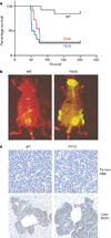
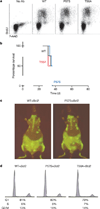
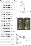
The c-Myc oncoprotein promotes proliferation and apoptosis, such that mutations that disable apoptotic programmes often cooperate with MYC during tumorigenesis. Here we report that two common mutant MYC alleles derived from human Burkitt's lymphoma uncouple proliferation from apoptosis and, as a result, are more effective than wild-type MYC at promoting B cell lymphomagenesis in mice. Mutant MYC proteins retain their ability to stimulate proliferation and activate p53, but are defective at promoting apoptosis due to a failure to induce the BH3-only protein Bim (a member of the B cell lymphoma 2 (Bcl2) family) and effectively inhibit Bcl2. Disruption of apoptosis through enforced expression of Bcl2, or loss of either Bim or p53 function, enables wild-type MYC to produce lymphomas as efficiently as mutant MYC. These data show how parallel apoptotic pathways act together to suppress MYC-induced transformation, and how mutant MYC proteins, by selectively disabling a p53-independent pathway, enable tumour cells to evade p53 action during lymphomagenesis.
Figures


Comment in
-
Cancer: two in one.Nature. 2005 Aug 11;436(7052):787-9. doi: 10.1038/436787a. Nature. 2005. PMID: 16094355 No abstract available.
Similar articles
-
Cancer: two in one.Nature. 2005 Aug 11;436(7052):787-9. doi: 10.1038/436787a. Nature. 2005. PMID: 16094355 No abstract available.
-
Bim is a suppressor of Myc-induced mouse B cell leukemia.Proc Natl Acad Sci U S A. 2004 Apr 20;101(16):6164-9. doi: 10.1073/pnas.0401471101. Epub 2004 Apr 12. Proc Natl Acad Sci U S A. 2004. PMID: 15079075 Free PMC article.
-
Bax loss impairs Myc-induced apoptosis and circumvents the selection of p53 mutations during Myc-mediated lymphomagenesis.Mol Cell Biol. 2001 Nov;21(22):7653-62. doi: 10.1128/MCB.21.22.7653-7662.2001. Mol Cell Biol. 2001. PMID: 11604501 Free PMC article.
-
Role of genetic and epigenetic changes in Burkitt lymphoma.Semin Cancer Biol. 2002 Oct;12(5):381-7. doi: 10.1016/s1044-579x(02)00058-5. Semin Cancer Biol. 2002. PMID: 12191637 Review.
-
EBV regulates c-MYC, apoptosis, and tumorigenicity in Burkitt's lymphoma.Curr Top Microbiol Immunol. 2001;258:153-60. doi: 10.1007/978-3-642-56515-1_10. Curr Top Microbiol Immunol. 2001. PMID: 11443860 Review. No abstract available.
Cited by
-
A critical role for Mnt in Myc-driven T-cell proliferation and oncogenesis.Proc Natl Acad Sci U S A. 2012 Nov 27;109(48):19685-90. doi: 10.1073/pnas.1206406109. Epub 2012 Nov 12. Proc Natl Acad Sci U S A. 2012. PMID: 23150551 Free PMC article.
-
MYC pathway activation in triple-negative breast cancer is synthetic lethal with CDK inhibition.J Exp Med. 2012 Apr 9;209(4):679-96. doi: 10.1084/jem.20111512. Epub 2012 Mar 19. J Exp Med. 2012. PMID: 22430491 Free PMC article.
-
A chemogenomic screening identifies CK2 as a target for pro-senescence therapy in PTEN-deficient tumours.Nat Commun. 2015 Jun 18;6:7227. doi: 10.1038/ncomms8227. Nat Commun. 2015. PMID: 26085373
-
Altering MYC phosphorylation in the epidermis increases the stem cell population and contributes to the development, progression, and metastasis of squamous cell carcinoma.Oncogenesis. 2020 Sep 7;9(9):79. doi: 10.1038/s41389-020-00261-3. Oncogenesis. 2020. PMID: 32895364 Free PMC article.
-
Global and context-specific transcriptional consequences of oncogenic Fbw7 mutations.Elife. 2022 Feb 28;11:e74338. doi: 10.7554/eLife.74338. Elife. 2022. PMID: 35225231 Free PMC article.
References
-
- Cole MD. Activation of the c-myc oncogene. Basic Life Sci. 1986;38:399–406. - PubMed
-
- Spencer CA, Groudine M. Control of c-myc regulation in normal and neoplastic cells. Adv. Cancer Res. 1991;56:1–48. - PubMed
-
- Davis M, Malcolm S, Rabbitts TH. Chromosome translocation can occur on either side of the c-myc oncogene in Burkitt lymphoma cells. Nature. 1984;308:286–288. - PubMed
Publication types
MeSH terms
Substances
Grants and funding
LinkOut - more resources
Full Text Sources
Other Literature Sources
Research Materials
Miscellaneous

