

Cytokine expression profile in human pancreatic carcinoma cells and in surgical specimens: implications for survival
- PMID: 16094523
- PMCID: PMC11031060
- DOI: 10.1007/s00262-005-0047-0
Cytokine expression profile in human pancreatic carcinoma cells and in surgical specimens: implications for survival
Abstract
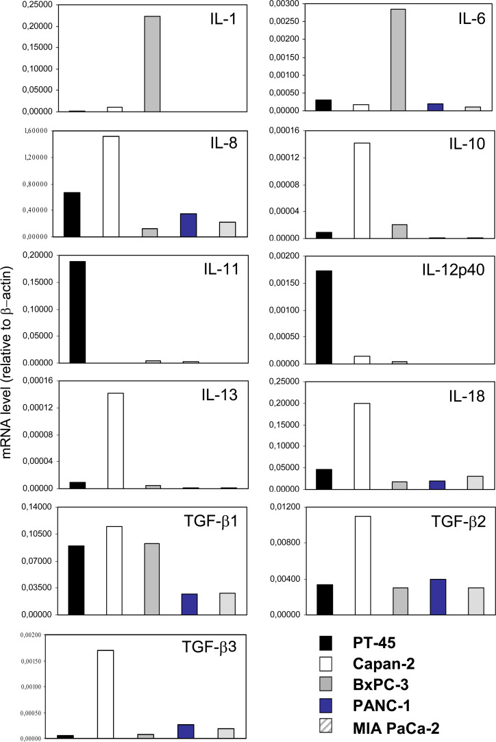
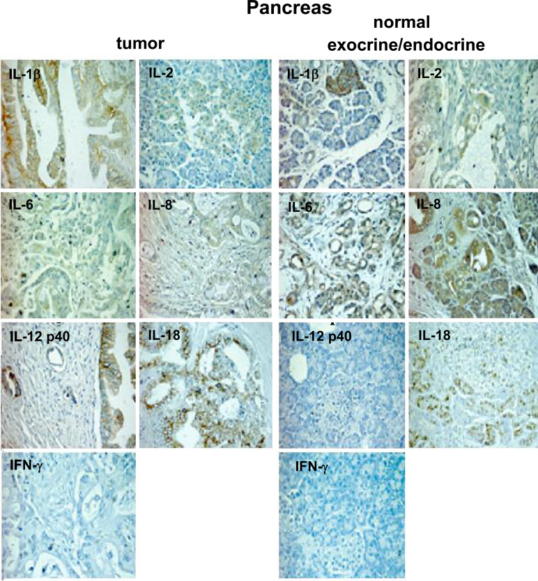
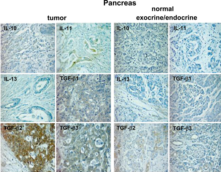
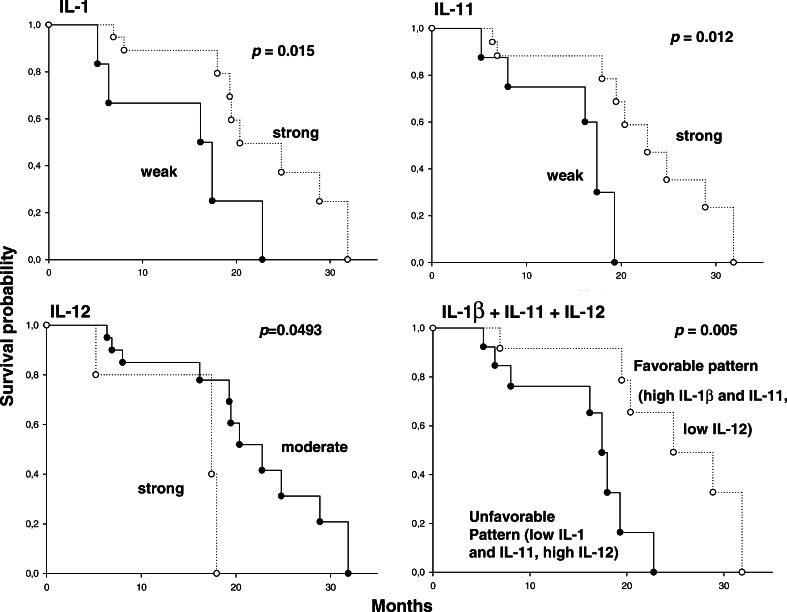
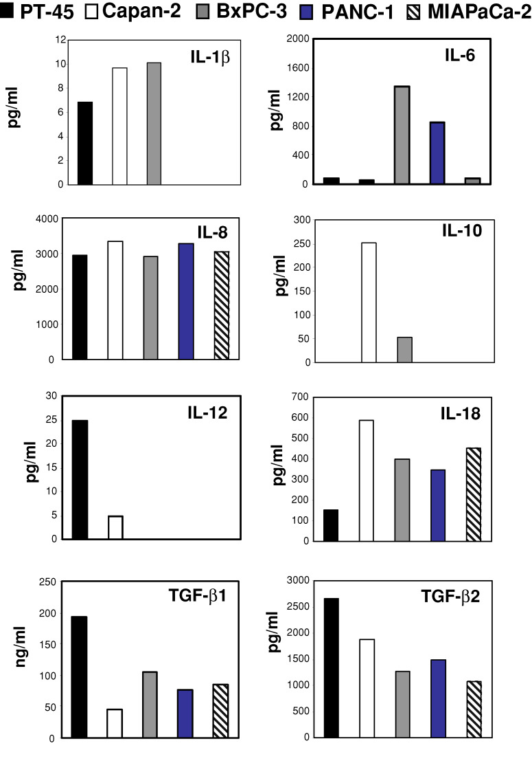
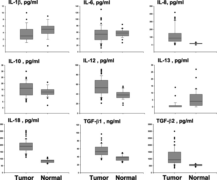
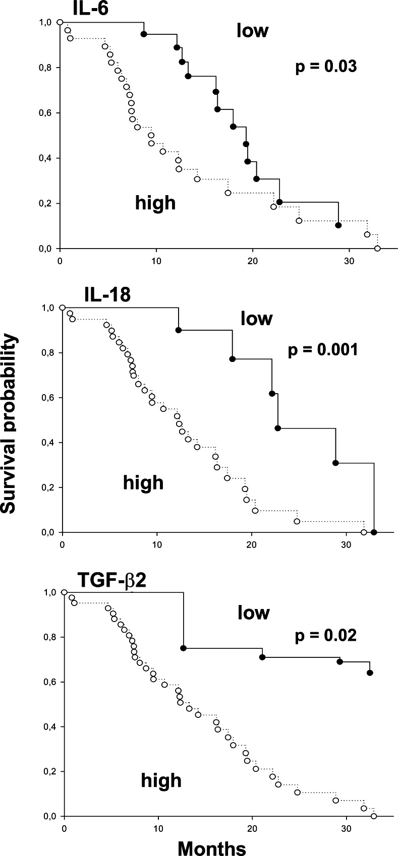
Cytokine shedding by tumor cells into the local microenvironment modulates host immune response, tumor growth, and metastasis. The study aimed to verify the hypothesis that the immunological microenvironment of pancreatic carcinoma exists in a prevalently immunosuppressive state, influencing survival. We analyzed expression profiles of pro-inflammatory (IL-1beta, IL-2, IL-6, IL-8, IL-12 p40, IL-18 and IFN-gamma) and anti-inflammatory (IL-10, IL-11, IL-13 and TGF-beta isoforms) cytokines. The study was performed both in vitro, in five pancreatic carcinoma cell lines (real time RT-PCR), and in specimens from 65 patients, comparing tumoral versus non-tumoral pancreatic tissues (real time RT-PCR and immunohistochemistry). Furthermore, cytokines were measured in supernatants and sera (from patients and controls) by ELISA. All cell lines expressed IL-8, IL-18, TGF-beta1, TGF-beta2 and TGF-beta3, but not IFN-gamma and IL-2 transcripts. Expression of IL-1beta, IL-6, IL-10, IL-11, IL-13 and IL-12 mRNA was variable. All the above cytokines were detected as soluble proteins in supernatants, except IL-13. Tumor tissues overexpressed IL-1beta, IL-6, IL-8, IL-10, IL-11, IL-12 p40, IL-18, IFN-gamma, TGF-beta1, TGF-beta2 and TGF-beta3 at the mRNA level and IL-1beta, IL-18, TGF-beta2 and TGF-beta3 also at the protein level. Conversely, non-tumor tissues had stronger RNA and protein expression of IL-13. Survival was significantly longer in patients with high IL-1beta and IL-11 and moderate IL-12 expression. Serum IL-8, IL-10, IL-12, IL-18, TGF-beta1 and TGF-beta2 were higher in patients than in controls, as opposed to IL-1beta and IL-13. Patients with low circulating levels of IL-6, IL-18 and TGF-beta2 survived longer. Pancreatic cancer is characterized by peculiar cytokine expression patterns, associated with different survival probabilities.
Figures
References
-
- Bellone G, Smirne C, Carbone A, Mareschi K, Dughera L, Farina EC, Alabiso O, Valente G, Emanuelli G, Rodeck U. Production and pro-apoptotic activity of soluble CD95 ligand in pancreatic carcinoma. Clin Cancer Res. 2000;6:2448–2455. - PubMed
Publication types
MeSH terms
Substances
LinkOut - more resources
Full Text Sources
Other Literature Sources
Medical
Miscellaneous
