

Mitral supravalvular ring: a case report
- PMID: 16095540
- PMCID: PMC1208923
- DOI: 10.1186/1476-7120-3-19
Mitral supravalvular ring: a case report
Abstract
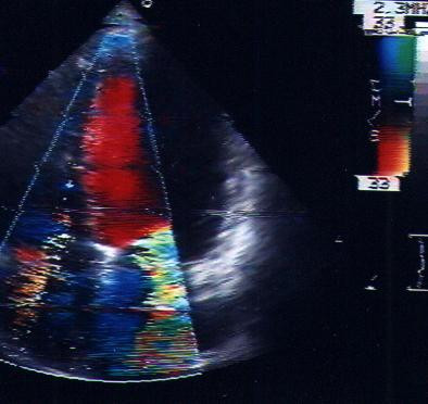
Supravalvular mitral stenosis is a rare condition characterized by an abnormal ridge, with one or two orifices, covering and obstructing the mitral valve. Preoperative diagnosis is difficult with transtoracic echo (TTE), angiography and magnetic resonance imaging (MRI). In this case, a 36-year-old male, was admitted to our Heart department: He experienced progressive dyspnea on effort and at rest. Diagnosis was made by transesophageal echocardiography which showed, on apical 4-chamber section, an annular structure attached since a membrane to the atrial wall anterior mitral valve leaflet and just proximal to the posterior mitral leaflet. Pre-operative identification of the supravalvular mitral ring is the target for obtaining good surgical results. Cineangiography and MRI both failed in reaching this objective, whereas, transesophageal echocardiography is the best method to identify this congenital heart disease. Using TEE the identification is not only possible but also easier.
Figures


References
-
- Lynch MF, Ryan NJ, William CR, Cayler G, Richardson WR, Campbell GS, Taybih Preoperative diagnosis and surgical correction of supravalvular mitral stenosis and ventricular septal defect. Circulation. 1962;25:85–61. - PubMed
-
- Coto EO, Judez VM, Juffe A, Rufilanchas JJ, Tellez G, Maronas J, Aymerich DF. Supravalvular stenotic mitral ring. A new case with surgical correction. J Thorac Cardiovasc Surg. 1976;71:537–539. - PubMed
-
- Maron BJ, Edwards JE, Ferrans VJ, Clark CE, Lebowitz EA, Henry WL, Epstein SE. Congenital heart malformations associated with disproportionate ventricular septal thickening. Circulation. 1975;52:926–932. - PubMed
-
- Mychaskiw G, 2nd, Sachdev V, Braden DA, Heath BJ. Supramitral ring: an unusual cause of congenital mitral stenosis. Case series and review. J Cardiovasc Surg (Torino) 2002;43:199–202. - PubMed
Publication types
MeSH terms
LinkOut - more resources
Full Text Sources
