. 2005 Oct 25;341(2):179-89.
doi: 10.1016/j.virol.2005.06.044. Epub 2005 Aug 10.

Homo-oligomerization facilitates the interferon-antagonist activity of the ebolavirus VP35 protein

Affiliations

Homo-oligomerization facilitates the interferon-antagonist activity of the ebolavirus VP35 protein

St Patrick Reid et al. Virology. .

Abstract

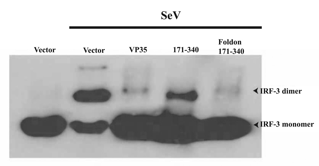
We have identified a putative coiled-coil motif within the amino-terminal half of the ebolavirus VP35 protein. Cross-linking studies demonstrated the ability of VP35 to form trimers, consistent with the presence of a functional coiled-coil motif. VP35 mutants lacking the coiled-coil motif or possessing a mutation designed to disrupt coiled-coil function were defective in oligomerization, as deduced by co-immunoprecipitation studies. VP35 inhibits signaling that activates interferon regulatory factor 3 (IRF-3) and inhibits (IFN)-alpha/beta production. Experiments comparing the ability of VP35 mutants to block IFN responses demonstrated that the VP35 amino-terminus, which retains the putative coiled-coil motif, was unable to inhibit IFN responses, whereas the VP35 carboxy-terminus weakly inhibited the activation of IFN responses. IFN-antagonist function was restored when a heterologous trimerization motif was fused to the carboxy-terminal half of VP35, suggesting that an oligomerization function at the amino-terminus facilitates an "IFN-antagonist" function exerted by the carboxy-terminal half of VP35.


Figures

Figure 1
Figure 1. VP35 forms oligomers
(A) Output from analysis of the Zaire ebolavirus VP35 protein by the COILS program. The X-axis represents the 340 amino acid VP35 sequence, and the Y-axis represents the probability (with 1=100%) that an amino acid residue is within a coiled-coil region. Different size scanning windows are indicated by different lines as shown in the inset. Window sizes are 14 (dashed line), 21 (dotted line) and 28 (solid line) residues. (B) Gel filtration analysis of VP35. Purified FLAG-VP35 was fractionated on a Superdex-200 fast-performance liquid chromatography column (Amersham Biosciences). Collected gel filtration fractions were analyzed by silver stain (top) and by western blot with anti-VP35 monoclonal antibody (bottom). The indicated fractions correspond to the following approximate molecular weights: fraction 17-194 kDa; 18-126 kDa; 19-81 kDa; and 20-52 kDa. Molecular weights were estimated by comparison to protein standards chymotrypsinogen A (19.9 kDa), bovine serum albumin (67 kDa), catalase (232 kDa), ferritin (440 kDa) and thyroglobulin (670 kDa), run under identical conditions. (C) DSP crosslinking analysis of VP35. Purified FLAG-VP35 was crosslinked with 0, 1, 2 or 5 mM DSP for 30 min and subsequently subjected to SDS-PAGE analysis under nonreducing conditions. The calculated molecular weights for each band are indicated by the arrows pointing to the corresponding band. Western blotting of gel filtration and crosslinking samples were performed using a monoclonal anti-VP35 antibody (6C5).
Figure 2
Figure 2. VP35-VP35 interaction requires the predicted coiled coil domain
(A) A schematic illustration of the wild-type and mutant forms of VP35 used for these experiments is presented. CCR, putative coiled-coil region. Foldon, foldon trimerization domain (see text). (B) 293T cells were transfected with expression plasmids encoding the indicated proteins. Twenty-four hours post-transfection cells were harvested and lysed. The prepared lysates were immunoprecipitated (IP) using anti-FLAG antibody (Sigma). After SDS-PAGE, western blotting was performed using anti-HA antibody (Sigma). Expression of HA and FLAG-tagged constructs were confirmed by Western blot analysis, with anti-HA and anti-FLAG tag antibodies, as is shown in the middle and lower panels. Note that oligomeric forms of wt VP35 and VP351–170 molecules are seen in the lower panel depicting the anti-HA western blot of total cell lysates.
Figure 3
Figure 3. Ability of full length and mutant VP35 constructs to inhibit activation of IRF-3 responsive genes
(A, B) Cells were co-transfected with empty expression plasmid or plasmids expressing the indicated wild-type or mutant VP35s along with an ISG54 promoter-driven CAT reporter gene and a constitutively expressed Renilla luciferase reporter plasmid. One day post-transfection, the cells were infected with SeV (moi=10) and, the following day, reporter gene activity was measured. Virus-induced ISG54 reporter values were normalized to the luciferase activity of a Renilla luciferase expressing plasmid. Results are presented as fold-induction of the ISG54 reporter relative to an empty vector-transfected, mock-infected control. In panel A, all dishes received 2.5 µg of expression plasmid. In panel B, 25 or 250 ng of expression plasmid (expressing empty vector (empty), full-length VP35 (VP35), VP35171–340 (171–340) or Foldon-VP35171–340) were transfected (concentrations indicated by wedges). For these experiments, empty vector was used to adjust all transfections to the same final DNA concentration. For both A and B, the error bars indicate standard deviation. In panel A, values not statistically different, as determined by a Student’s t-test (P≤ 0.05), are indicated by identical symbols (* and #). In panel B, the values for empty vector (empty)- and the 171–340 (lower 25 ng plasmid concentration)-transfected samples are statistically different from each other and all other samples, as determined by Student’s t-test (P≤ 0.05). (C) Western blot analysis of the indicated expression plasmids is provided (following transfection of 250 ng or 25 ng). The relative expression levels of these proteins are highly reproducible. (D) Expression of HA-VP35171–340 and Foldon-HA-VP35171–340 to verify trimer formation of Foldon-VP35171–340 construct. Lysates from cells transfected with the indicated construct were subjected western blot analysis with anti- HA antibody (Sigma). These samples were not boiled prior to electrophoresis.
Figure 4
Figure 4. An interferon bioassay demonstrates that the foldon motif restores to VP35171–340 the ability to inhibit endogenous IFNβ production
293T cells were transfected with the indicated amounts (left of the figure) of expression plasmids encoding VP35, VP35171–340, and Foldon-VP35171–340. The total amount of expression plasmid in all transfections was adjusted to a 2500 ng with empty vector. As a control, cells were transfected with 2500 ng empty vector (top two panels). 24 hours post-transfection, the 293T cells were mock infected or infected with SeV (moi=10). One day post-infection media supernatants from all cultures were collected, clarified by centrifugation and UV treated to inactivate any infectious SeV. A. To test for IFN antiviral activity, the inactivated supernatants were transferred onto Vero cells at the indicated dilutions (1:256 or 1:512, indicated by “Reciprocal of dilution”). The treated Vero cells were subsequently infected with the NDV-GFP virus. IFN present in the 293T cell supernatants, induced by the prior SeV infection, inhibits replication of NDV-GFP and green fluorescence in the Vero cells, (see the two uppermost panels, empty vector (2500 ng)). As the supernatnants are diluted, an increase in GFP expression is seen. In the anti-IFNβ panel, supernatant from an empty vector-transfected, SeV-infected cells was pre-incubated with IFNβ neutralizing antiserum prior to its addition to the Vero cells. The recovery of NDV-GFP replication in these cells demonstrates that the antiviral effect in these cells is mediated mainly by IFNβ. The “Mock infected” control panel denotes Vero cells without NDV-GFP infection. VP35, 171–340 and Foldon 171–340 indicate the expression plasmids transfected at the indicated amount (in ng/106 cells) (see Expression vector amounts at left). B. To assess SeV-induced expression of the IRF-3-responsive ISG56 gene, western blots were performed to examine levels of p56, the protein product of the ISG56 gene. Expression of p56, 1 day post SeV-infection of cells tranfected with 25, 250 or 2500 ng of the indicated expression plasmids, is shown.
Figure 5
Figure 5. The foldon trimerization motif restores the ability of VP35171–340 to block IRF-3 activation
293T cells were transfected with the indicated protein expression plasmids (full length VP35 (VP35), VP35171–340 (171–340) or Foldon-VP35171–340 (Foldon 171–340) and either mock-infected of infected with Sendai virus (SeV) 24hr post-transfection. Ten hours post-infection cells were harvested and lysed. For IRF-3 analysis, cells were subjected to native PAGE analysis as previously described (Iwamura reference) followed by blotting with an anti- IRF-3 monoclonal antibody.

Similar articles

Cited by

References

    1. Andrejeva J, Childs KS, Young DF, Carlos TS, Stock N, Goodbourn S, Randall RE. The V proteins of paramyxoviruses bind the IFN-inducible RNA helicase, mda-5, and inhibit its activation of the IFN-beta promoter. Proc Natl Acad Sci U S A. 2004;101(49):17264–17269. - PMC - PubMed
    1. Basler CF, Mikulasova A, Martinez-Sobrido L, Paragas J, Muhlberger E, Bray M, Klenk HD, Palese P, Garcia-Sastre A. The Ebola virus VP35 protein inhibits activation of interferon regulatory factor 3. J Virol. 2003;77(14):7945–7956. - PMC - PubMed
    1. Basler CF, Wang X, Muhlberger E, Volchkov V, Paragas J, Klenk HD, Garcia-Sastre A, Palese P. The Ebola virus VP35 protein functions as a type I IFN antagonist. Proc Natl Acad Sci U S A. 2000;97(22):12289–12294. - PMC - PubMed
    1. Bosio CM, Aman MJ, Grogan C, Hogan R, Ruthel G, Negley D, Mohamadzadeh M, Bavari S, Schmaljohn A. Ebola and Marburg viruses replicate in monocytederived dendritic cells without inducing the production of cytokines and full maturation. J Infect Dis. 2003;188(11):1630–1638. - PubMed
    1. Bossert B, Conzelmann KK. Respiratory syncytial virus (RSV) nonstructural (NS) proteins as host range determinants: a chimeric bovine RSV with NS genes from human RSV is attenuated in interferon-competent bovine cells. J Virol. 2002;76(9):4287–4293. - PMC - PubMed

Publication types

MeSH terms