. 2005 Aug 12;122(3):379-91.
doi: 10.1016/j.cell.2005.06.015.

Escaping the nuclear confines: signal-dependent pre-mRNA splicing in anucleate platelets

Affiliations

Escaping the nuclear confines: signal-dependent pre-mRNA splicing in anucleate platelets

Melvin M Denis et al. Cell. .

Abstract

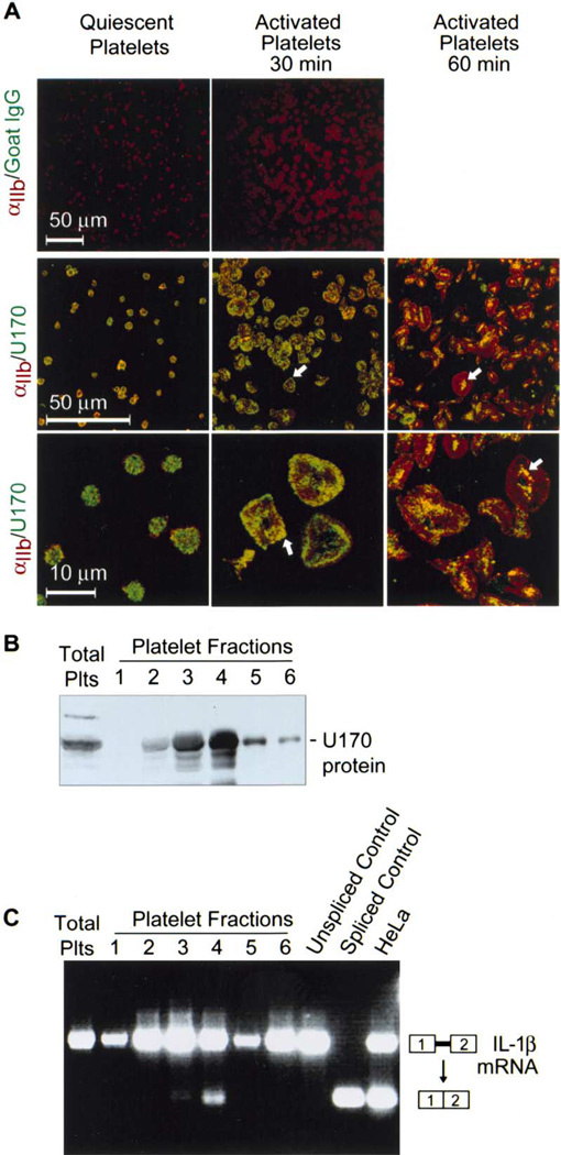
Platelets are specialized hemostatic cells that circulate in the blood as anucleate cytoplasts. We report that platelets unexpectedly possess a functional spliceosome, a complex that processes pre-mRNAs in the nuclei of other cell types. Spliceosome components are present in the cytoplasm of human megakaryocytes and in proplatelets that extend from megakaryocytes. Primary human platelets also contain essential spliceosome factors including small nuclear RNAs, splicing proteins, and endogenous pre-mRNAs. In response to integrin engagement and surface receptor activation, platelets precisely excise introns from interleukin-1beta pre-mRNA, yielding a mature message that is translated into protein. Signal-dependent splicing is a novel function of platelets that demonstrates remarkable specialization in the regulatory repertoire of this anucleate cell. While this mechanism may be unique to platelets, it also suggests previously unrecognized diversity regarding the functional roles of the spliceosome in eukaryotic cells.


Figures

Figure 1
Figure 1. U1 70K Protein Is Localized in the Cytoplasm of Megakaryocytes and in Pro-platelets that Extend from Megakaryocytes
(A) Scanning electron micrographs of a CD34+ stem-cell-derived megakaryocyte (left panel) and a megakaryocyte with proplatelet extensions (right panel). The white arrows in the right panel point to proplatelets, while the blue arrow indicates the cell body. (B) Transmission electron micrographs of a megakaryocyte (top left panel) and a megakaryocyte with a proplatelet extension (top right panel). The arrow in the top panel is pointing to the proplatelet extension. The bottom left panel is a corresponding photomicrograph of the megakaryocyte taken at a higher magnification to identify the nuclear membrane. The bottom middle and right panels are corresponding photomicrographs of the megakaryocyte with a proplatelet extension taken at higher magnifications to identify the nuclear membrane. The black arrows in the bottom panels identify the nuclear envelope. (C) A representative megakaryocyte and a megakaryocyte with proplatelet extensions were costained with an anti-U1 70K antibody (green) and wheat germ agglutinin (WGA; red), a fluorescent lectin that binds cell membranes and granules. In the megakaryocyte, U1 70K protein is detected in the cytoplasm (white arrow in the top, right panel) as well as in the nucleus. U1 70K protein is also observed in proplatelet extensions of the megakaryocyte (white arrows in the bottom, right panel). The right panels are merged images of WGA and U1 70K protein. The blue arrow in the bottom panel indicates U1 70K staining in the nucleus. The photomicrographs in this figure are representative of three independent experiments.
Figure 2
Figure 2. Critical Spliceosome Factors Are Present in the Cytoplasm of Megakaryocytes, in Megakaryocytes with Proplatelet Extensions, and in Circulating Platelets
(A and B) Megakaryocytes were stained with an anti-U2AF65 (A) or anti-SF2/ASF (B) and an antibody against the integrin αIIb subunit. Low-magnification photomicrographs for each factor are shown in grayscale. The merged photomicrographs in the right panels are higher magnifications of the areas outlined in blue in the middle panels. In the right panels, U2AF65 and SF2/ASF immuno-localization is shown in green, and αIIb immunostaining is in red. The white arrows in the top panels indicate cytoplasmic staining and in the bottom panels identify immunostaining in proplatelet extensions. (C) Platelets were left in suspension (quiescent) or allowed to adhere and spread on immobilized fibrinogen in the presence of thrombin for 30 or 60 min. The platelets were stained with antibodies against U2AF65 (top panels) or SF2/ASF (bottom panels) and the αIIb integrin subunit (U2AF65 and SF2/ASF immunolocalization, green; αIIb immunostaining, red). Controls for antibody specificity were performed (see Figure 3C for examples). Cellular spreading on immobilized fibrinogen in the presence of thrombin demonstrates that the platelets were efficiently stimulated. U2AF65 and SF2/ASF were also detected in platelets by Western blotting (data not shown). The images in this figure are representative of three independent studies.
Figure 3
Figure 3. Localization of U snRNAs in Megakaryocytes, Megakaryocytes with Proplatelet Extensions, and Circulating Platelets
(A) Localization of U snRNAs in a megakaryocyte (left panel) and a megakaryocyte with a proplatelet extension (right panel) was characterized with an anti-TMG antibody. Staining of U snRNAs and the αIIb integrin subunit are shown in green and red, respectively (blue arrows, nuclear localization of U snRNAs; white arrows, cytoplasmic localization of U snRNAs). (B) Northern analysis of U snRNAs (U1, U2, U4, U5, and U6) in HeLa cells and circulating platelets (Plt). The two panels on the right are Northern blots conducted in the presence of competing unlabeled oligonucleotides demonstrating the specificity of the U snRNA probes. (C) The top panels are control studies of circulating platelets (left and middle panels) and HeLa cells (right panel), respectively. In the platelet controls, the cells were incubated with nonimmune mouse IgG and a specific antibody against the αIIb integrin subunit (red stain). The HeLa cells (top right panel) were costained with anti-TMG (green stain) and phalloidin (red stain), a marker specific for polymerized actin. In the bottom panels, an anti-TMG antibody (green) was used to localize U snRNAs in quiescent platelets (bottom left panel) and platelets adherent to immobilized fibrinogen in the presence of thrombin for 30 (bottom middle panel) or 60 (bottom right panel) min. Staining for αIIb is in red. Each panel in this figure is representative of three independent experiments.
Figure 4
Figure 4. IL-1β Pre-mRNA Is Present in Developing Proplatelets as well as Quiescent Platelets, and Mature IL-1β mRNA Is Present in Activated Platelets
(A) In situ PCR for intronic IL-1β mRNA was conducted as described in detail in the Experimental Procedures. The top panels illustrate assays using probes for introns 1, 4, and 6 and demonstrate that IL-1β pre-mRNA is present in the cytoplasm of CD34+-derived megakaryocytes that are in the process of extending proplatelets. The black arrows identify cytoplasm of the megakaryocytes. The bottom panels illustrate corresponding negative controls in which reverse transcriptase was eliminated from the RT reaction (No RT). Here, the black arrows point to unstained megakaryocytes. (B) In situ PCR for IL-1β pre-mRNA and mature mRNA was conducted in quiescent platelets (top left panel) and in platelets adherent to fibrinogen in the presence of thrombin for 1 hr (top middle and right panels), respectively. The far right photomicrographs of adherent, activated platelets are taken at a higher magnification. In the bottom panels (No RT), the reverse transcriptase was omitted during the RT reaction. This figure represents inspection of multiple fields from three independent experiments.
Figure 5
Figure 5. Activated Platelets Splice Endogenous IL-1β Pre-mRNA into Mature Message and Translate the mRNA into Protein
(A) mRNA levels in platelets that were left quiescent (lane 1) or adhered to fibrinogen in the presence of thrombin (lanes 2–4). Lane 5 identifies mRNA levels in the CD45+ fraction extracted from this platelet preparation. On the right-hand side, the boxes represent exon 1 and 2 of the IL-1β message, which flank intron 1 (solid line). (B) In the left panel, mRNA from platelets that were left quiescent (0) or activated by fibrinogen and thrombin for 30 min was left unmanipulated (control) or treated with RNase or DNase. The right panel depicts genomic DNA treated with DNase and RNase. (C) Analysis of IL-1β pre-mRNA and mature mRNA in quiescent platelets and in platelets activated by adherence to fibrinogen for 2 hr in the presence of thrombin. The IL-1β gene is depicted in the top portion of this figure where exon flanking primer sets are color coded to indicate the approximate location of individual PCR reactions that span each intron of the IL-1β gene. On the right side, the boxes represent undesignated exons flanking a representative intron to illustrate the patterns of PCR products. (D) Polymerized actin (red) and IL-1β protein (green) were stained in quiescent platelets and in platelets 8 hr after they adhered to fibrinogen in the presence of thrombin (Activated). IL-1β protein was detected in adherent platelets consistent with de novo synthesis of the protein by platelets that are activated by fibrinogen and thrombin (Lindemann et al., 2001a).
Figure 6
Figure 6. Platelet Extracts Splice In Vitro-Transcribed IL-1β pre-mRNA
(A) This panel demonstrates immunolocalization of U1 70K protein in mature platelets. In the top panels, fixed platelets were incubated with nonimmune goat IgG and the antibody against the αIIb integrin subunit (red stain). In the subsequent panels, low-magnification (middle row) and high-magnification (bottom row) photomicrographs illustrate platelets that were costained with antibodies directed against U1 70K protein (green) and αIIb (red). Each panel is representative of six independent experiments. (B) Platelet lysates were left unfractionated (total plts) or were separated into six fractions on sucrose gradients. U1 70K protein was detected by Western analysis. (C) In vitro-transcribed IL-1β pre-mRNA was incubated with unfractionated platelet lysates (total plts), fractions 1–6, without platelet lysates (unspliced control) or with HeLa cell nuclear extracts (last lane). In vitro-transcribed intronless IL-1β mRNA (spliced control) was analyzed on the same gel as a positive control for spliced message. Gels in Figures 6B and 6C are representative of three independent experiments.

Comment in

  • Splicing misplaced.
    Meshorer E, Misteli T. Meshorer E, et al. Cell. 2005 Aug 12;122(3):317-8. doi: 10.1016/j.cell.2005.07.016. Cell. 2005. PMID: 16096050 Review.

References

    1. Albertine KH, Jones GP, Starcher BC, Bohnsack JF, Davis PL, Cho SC, Carlton DP, Bland RD. Chronic lung injury in preterm lambs. Disordered respiratory tract development. Am. J. Respir. Crit. Care Med. 1999;159:945–958. - PubMed
    1. Bahou WF, Gnatenko DV. Platelet transcriptome: the application of microarray analysis to platelets. Semin. Thromb. Hemost. 2004;30:473–484. - PubMed
    1. Black DL. Mechanisms of alternative pre-messenger RNA splicing. Annu. Rev. Biochem. 2003;72:291–336. - PubMed
    1. Blencowe BJ, Issner R, Nickerson JA, Sharp PA. A coactivator of pre-mRNA splicing. Genes Dev. 1998;12:996–1009. - PMC - PubMed
    1. Brogren H, Karlsson L, Andersson M, Wang L, Erlinge D, Jern S. Platelets synthesize large amounts of active plasminogen activator inhibitor 1. Blood. 2004;104:3943–3948. - PubMed

Publication types

MeSH terms