. 2005 Sep 7;24(17):3082-92.
doi: 10.1038/sj.emboj.7600772. Epub 2005 Aug 11.

Prion generation in vitro: amyloid of Ure2p is infectious

Affiliations

Prion generation in vitro: amyloid of Ure2p is infectious

Andreas Brachmann et al. EMBO J. .

Abstract

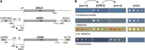
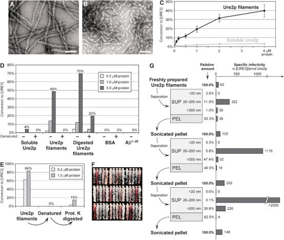
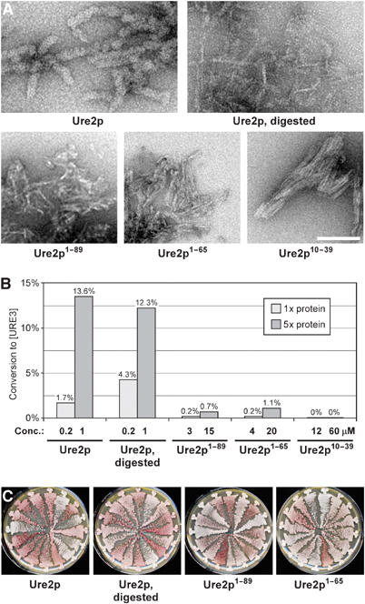
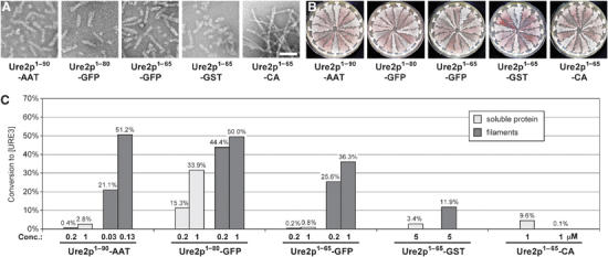
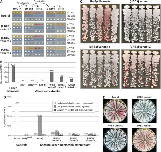
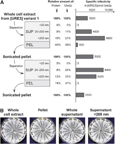
[URE3] is a prion (infectious protein) of the Ure2 protein of yeast. In vitro, Ure2p can form amyloid filaments, but direct evidence that these filaments constitute the infectious form is still missing. Here we demonstrate that recombinant Ure2p converted into amyloid can infect yeast cells lacking the prion. Infection produced a variety of [URE3] variants. Extracts of [URE3] strains, as well as amyloid of Ure2p formed in an extract-primed reaction could transmit to uninfected cells the [URE3] variant present in the cells from which the extracts were made. Infectivity and determinant of [URE3] variants resided within the N-terminal 65 amino acids of Ure2p. Notably, we could show that amyloid filaments of recombinant Ure2p are nearly as infectious per mass of Ure2p as extracts of [URE3] strains. Sizing experiments indicated that infectious particles in vitro and in vivo were >20 nm in diameter, suggesting that they were amyloid filaments and not smaller oligomeric structures. Our data indicate that there is no substantial difference between filaments formed in vivo and in vitro.


Figures

Figure 1
Figure 1
Assays for [URE3]. Ure2p negatively regulates the DAL5 promoter in the presence of a rich nitrogen source. Inactivation of Ure2p, either by prion formation, or in ure2Δ strains, leads to transcription from the DAL5 promoter. DAL5 encodes the allantoate transporter, which also takes up ureidosuccinate, the product of Ura2p (aspartate transcarbamylase). In this study, the DAL5 promoter was placed upstream of the ADE2 and CAN1 open reading frames. In [URE3] or ure2Δ strains containing these constructs, a derepressed DAL5 promoter allowed growth of a ura2 mutant when ureidosuccinate was substituted for uracil. Adenine prototrophy, white colony color on limiting adenine, and sensitivity to the arginine analog canavanine taken up by Can1p can be observed. (A) Schematic representation of the genomic DAL5, ADE2, and CAN1 loci in the reporter strains. Dotted lines indicate the deleted promoter regions. (B) BY334 cells lacking [URE3] (‘[ure-o]') were converted to [URE3] by cytoduction from strain 3310 (‘[URE3]'). Those cytoductants were cured of [URE3] by growth for 3 days on 1/2 YPD plates containing 3 mM guanidine (‘[ure-o]'). ure2Δ control strains were 4132 and BY256. Serial 10-fold dilutions were spotted on SD+Ade,HLW plates containing 33 mg/l ureidosuccinate (‘+Ureidosuccinate'), on SC−Ade plates (‘−Adenine'), on 1/2 YPD plates (‘Low adenine'), and on HC−R plates containing 200 mg/l canavanine (‘+Canavanine').
Figure 2
Figure 2
Transformation of in vitro-formed Ure2p filaments into yeast spheroplasts. (A) Electron micrograph of negatively stained Ure2p filaments. Bar, 100 nm. (B) Negatively stained Ure2p filaments after sonication two times for 15 s at 60 W. Bar, 100 nm. (C) Ure2p concentration dependence of conversion to [URE3]. Indicated concentrations of Ure2p filaments and soluble Ure2p were transformed into BY241. Values of [URE3] colonies relative to all Leu+ transformants are the mean of at least three independent experiments (±standard error). (D) Infectivity of proteinase K-digested Ure2p filaments (see Materials and methods) and effect of heat denaturation. Heat denaturation of protein samples was achieved by incubation at 95°C for 5 min. Indicated protein concentrations were transformed into BY241. (E) Recovery of infectivity from heat-denatured Ure2p filaments by proteinase K digestion. Heat denaturation, proteinase K treatment, and transformation were performed as in panel D. (F) Spectrum of [URE3] variants in randomly chosen colonies after transformation of BY241 with Ure2p filaments. (G) Fractionation of freshly prepared Ure2p filaments (see Materials and methods) and specific infectivity following transformation into BY241. The starting concentration of Ure2p filaments was 2 μM; concentrations of each fraction are presented as relative values. Electron micrographs of filament fractions and [URE3] variant spectra of the respective transformants are provided in Supplementary Figure 3.
Figure 3
Figure 3
Transformation of filaments from Ure2p N-terminal fragments. (A) Negatively stained filaments of the indicated proteins. Bar, 100 nm. (B) Infectivity of filaments from Ure2p N-terminal fragments in comparison to full-length Ure2p filaments and proteinase K-digested Ure2p filaments. Indicated protein concentrations were transformed into BY241. (C) Spectrum of [URE3] variants in randomly chosen transformants obtained from the successful transformations in panel B.
Figure 4
Figure 4
Transformation of fusion protein filaments. (A) Negatively stained filaments of the indicated proteins. Bar, 100 nm. (B) Spectrum of [URE3] variants resulting from transformation of BY241 with fusion protein filaments. In the case of Ure2p1−65-CA, transformants were obtained from transformation of soluble protein. (C) Infectivity of soluble protein and filaments from fusion proteins, as determined by transformation of strain BY241 with the indicated concentrations of protein.
Figure 5
Figure 5
Transformation of [URE3] variants. (A) Conservation of variant characteristics upon transmission of [URE3] variants. The cytoplasm of BY241 strains that were [ure-o] or one of the three different [URE3] variants was transferred to BY251 by cytoduction, and then cytoduced back into BY241. Recipient clones were cured of [URE3] by growth on 1/2 YPD plates containing 3 mM guanidine, and demonstrated reversion to [ure-o] in all cases. Serial 10-fold dilutions were spotted on the indicated media and incubated at 30°C for 3 days (1/2 YPD and SC+Can) or 5 days (SC−Ade). Note the differences in color on 1/2 YPD plates and sensitivity to canavanine between the three [URE3] variants and between the two strains. Data from a parallel experiment performed with BY252 instead of BY251 are provided in Supplementary Figure 5. (B) Infectivity of whole cell extracts from strains BY259 (ure2fs), BY277 (URE290354), and BY241 with [ure-o] or one of the three [URE3] variants, as determined by transformation into BY241. Protein extracts were transformed at the following concentrations (in mg/ml): ure2fs, 0.5; URE290354, 0.8; [ure-o], 1.3; [URE3] variant 1, 1.1; [URE3] variant 2, 1.3; [URE3] variant 3, 1.3. A control transformation was performed with 10 μM Ure2p filaments. Control transformations to ensure that the obtained [URE3] clones are indeed transformants and not cells remaining in the extracts are described in Supplementary data. (C) Spectrum of [URE3] variants following successful transformations in panel B. Five randomly chosen clones were streaked on 1/2 YPD plates and incubated at 30°C for 5 days. (D) Seeding experiment with whole cell extracts. Solutions of Ure2p and Ure2p90−354 were incubated with or without agitation (on a vertical roller at 10 r.p.m.) at 4°C for 12 h with whole cell extracts from BY241 strains that were [ure-o] or contained one of the three [URE3] variants. The Ure2p to extract ratio was roughly 1/150 and corresponds to a Ure2p ratio of about 1/2 × 106. The solutions were sonicated and transformed into BY241 at a concentration of 2 μM. For comparison, soluble Ure2p and Ure2p90−354 were incubated in the same way and also transformed into BY241. (E) Spectrum of [URE3] variants following transformation of agitated seeding solutions in panel D.
Figure 6
Figure 6
Fractionation of whole cell extracts. (A) Cellular extract of strain BY251 [URE3] variant 1 was fractionated (see Materials and methods) and transformed into BY241. The starting concentration was 25 mg/ml; relative amounts of Ure2p were estimated by determining whole protein concentration and relative Ure2p content in each fraction (see Materials and methods). Similar results were obtained with cell extracts from different strains and [URE3] variants (data not shown). (B) Fractionation does not interfere with faithful transmission of [URE3] characteristics to the transformants. All randomly chosen transformants resulting from a transformation with BY241 [URE3] variant 1 extract fractions show the characteristic white appearance on 1/2 YPD plates. Extract fractionation was performed as in previous experiments, except that pelleting was performed at 45 000 g for 45 min.

References

    1. Aigle M, Lacroute F (1975) Genetical aspects of [URE3], a non-Mendelian, cytoplasmically inherited mutation in yeast. Mol Gen Genet 136: 327–335 - PubMed
    1. Amberg D, Burke DJ, Strathern JN (2005) Methods in Yeast Genetics. Woodbury, NY, USA: Cold Spring Harbor Laboratory Press
    1. Baxa U, Cheng N, Winkler DC, Chiu TK, Davies DR, Sharma D, Inouye H, Kirschner DA, Wickner RB, Steven AC (2005) Filaments of the Ure2p prion protein have a cross-beta core structure. J Struct Biol 150: 170–179 - PubMed
    1. Baxa U, Speransky V, Steven AC, Wickner RB (2002) Mechanism of inactivation on prion conversion of the Saccharomyces cerevisiae Ure2 protein. Proc Natl Acad Sci USA 99: 5253–5260 - PMC - PubMed
    1. Baxa U, Taylor KL, Wall JS, Simon MN, Cheng N, Wickner RB, Steven A (2003) Architecture of Ure2p prion filaments: the N-terminal domain forms a central core fiber. J Biol Chem 278: 43717–43727 - PubMed

MeSH terms