. 2005 Sep 7;24(17):3070-81.
doi: 10.1038/sj.emboj.7600770. Epub 2005 Aug 11.

Acetylation of HIV-1 integrase by p300 regulates viral integration

Affiliations

Acetylation of HIV-1 integrase by p300 regulates viral integration

Anna Cereseto et al. EMBO J. .

Abstract

Integration of HIV-1 into the human genome, which is catalyzed by the viral protein integrase (IN), preferentially occurs near transcriptionally active genes. Here we show that p300, a cellular acetyltransferase that regulates chromatin conformation through the acetylation of histones, also acetylates IN and controls its activity. We have found that p300 directly binds IN both in vitro and in the cells, as also specifically demonstrated by fluorescence resonance energy transfer technique analysis. This interaction results in the acetylation of three specific lysines (K264, K266, K273) in the carboxy-terminus of IN, a region that is required for DNA binding. Acetylation increases IN affinity to DNA, and promotes the DNA strand transfer activity of the protein. In the context of the viral replication cycle, point mutations in the IN acetylation sites abolish virus replication by specifically impairing its integration capacity. This is the first demonstration that HIV-1 IN activity is specifically regulated by post-translational modification.


Figures

Figure 1
Figure 1
HIV-1 IN binds p300 in vitro and in vivo. (A) IN interacts with p300 in vivo. Extracts prepared from cells transfected with pFlag-IN or pFlag-Luc vectors were immunoprecipitated with an anti-p300 antibody and immunoblotted with an anti-Flag antibody (upper panel) or an anti-p300 antibody (lower panel). The same extracts were run on an SDS–PAGE gel and immunoblotted with an anti-Flag antibody (right panel). (B) Extracts as in (A) were immunoprecipitated with anti-Flag antibody and immunoblotted with anti-p300 antibody (upper panel). The same extracts were analyzed by immunoblotting with an anti-p300 antibody (lower panel). (C) In vitro IN/p300 binding. Serial dilutions of GST-IN on agarose beads were incubated with fixed amounts of 35S-p300 and analyzed on an SDS–PAGE gel. (D) GST-IN fragments were incubated with 35S-p300 and analyzed on an SDS–PAGE gel. In (C) and (D), the upper panels show the Coomassie staining of the gels and the lower panel gels exposed to Cyclone screen. The graphs express the amounts of bound proteins as percentages of the input of radiolabeled protein.
Figure 2
Figure 2
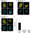
FRET analysis of protein–protein interaction. (A) Visualization of FRET. pEYFP-IN and pECFP-p300 vectors were transfected in U2OS cells and visualized by excitation at 514 nm showing EYFP fluorescence (upper panels), or at 458 nm showing ECFP fluorescence (lower panels). In the upper left panel, the region chosen for photobleaching (ROI) is indicated. (B) As in (A), following transfection with pEYFP-ΔIN 1–263 and pECFP-p300. (C) As in (A), following transfection with pEYFP-CDK9 and pECFP-Rb. (D) Quantification of FRET. The FRET efficiency was measured by comparing the fluorescence intensity of the donor ECFP before and after photobleaching of the acceptor EYFP within the ROI (see Materials and methods). Bar scale: 5 μm.
Figure 3
Figure 3
IN is acetylated by p300 both in vitro and in vivo. (A) In vitro IN acetylation. p300-HAT was incubated with GST-IN (lane 3) or controls (lanes 1 and 2) in the presence of 14C acetyl-CoA; after incubation, the reaction mixture was resolved by SDS–PAGE and the gel exposed to Cyclone screen. Lanes 4–6 show Coomassie blue staining of the same gel. (B) Schematic representation of the GST-IN proteins used for pulldown and acetylation assays. The positions of the lysines in the IN C-terminal domain are indicated. The lysines positive for acetylation are shown in red. (C) The IN full-length protein (lane 2), the N-terminal, core, and C-terminal domains (lanes 3–5), a series of IN mutants with C-terminal truncations (lanes 6–9), and point mutants in the lysines in the C-terminal domain (lanes 12–18) were assayed for acetylation by p300-HAT. Upper panels: gels exposed to Cyclone screen. Lower panels: Coomassie-stained gels. (D) In vivo IN acetylation. Extracts of 293T cells transfected as indicated were immunoprecipitated with an anti-Flag antibody and immunoblotted using an anti-Ac-Lys antibody (upper panels). The same filters were incubated with an anti-Flag antibody (lower panels).
Figure 4
Figure 4
Acetylation of IN increases DNA binding. (A) Scalar amounts of His-IN were preincubated with p300 HAT either in the absence (lanes 1–5) or presence (lane 6–10) of cold acetyl-CoA, followed by UV-crosslinking DNA-binding analysis (see Materials and methods). The bands correspond to the complex between probe and IN. (B) The graph summarizes the results obtained from three independent experiments performed as in panel A; the average and s.d. of the amount of probe bound to IN are shown. To compare the different experiments, the c.p.m. values were normalized to the amount of probe bound by the lowest amount of IN in the absence of acetyl-CoA. (C) Scalar amounts of His-IN either wt (lanes 1–9) or mutated in K(264,266,273)R (lanes 10–18) were preincubated with p300-HAT either in the absence (lanes 1–3 and 10–12) or presence of cold acetyl-CoA (lanes 4–9 and 13–18) in addition to Lys-CoA (lanes 7–9 and 16–18), followed by UV-crosslinking DNA-binding analysis (see Materials and methods). (D) The graph summarizes the results obtained from three independent experiments performed as in panel (C) and indicated as in (B). (E) HAT assay performed with recombinant His-IN wt (lanes 1–3) or mutated in K(264,266,273)R (lanes 4–6) in the presence of 14C acetyl-CoA (lanes 2, 3 and 5, 6) in addition to Lys-CoA (lanes 3 and 6).
Figure 5
Figure 5
Acetylation increases strand transfer activity of IN. (A) Strand transfer activity of His-IN wt (lanes 1, 2) or mutated in K(264,266,273)R (lanes 3, 4) preincubated with p300-HAT either in the absence (lanes 1, 3) or in the presence (lanes 2, 4) of cold acetyl-CoA. Lane 5: substrate without His-IN. (B) Quantification analysis of the strand transfer activity obtained from three independent experiments performed as in (A); the average and s.d. of the summary of the intensity of bands that result as products of the strand transfer activity in each lane are shown. (C) 3′ processing activity of His-IN, after preincubation with p300-HAT either in the absence (lane 1) or presence (lane 2) of cold acetyl-CoA. Lane 3: substrate without His-IN. In (A) and (C), the DNA substrate (S) and catalytic products (P) are indicated.
Figure 6
Figure 6
Replication of an HIV-1 viral clone carrying the IN K(264,266,273)R mutation is impaired. (A) Replication kinetics of wt HIV-1BRU (HIV wt) and of an HIV-1BRU viral clone carrying the IN K(264,266,273)R mutation (HIV mut). In both viral clones, the IN is Flag-tagged. CEM T-cells and PBLs from two healthy donors were infected with equal amounts (0.5 × 10⁷ c.p.m. RT) of the two viruses; the RT activity of the supernatants was measured at different time points after infection. Each graph shows the average values from three independent experiments. (B) RT activity of the supernatants measured at different time points after infection of CEM T cells with 1 × 10⁷ or 5 × 10⁷ c.p.m. RT of HIV wt or HIV mut. (C) Western blot analysis of either HIV wt or HIV mut viral stocks using an anti-MA (left panel) or an anti-Flag (right panel) antibody.
Figure 7
Figure 7
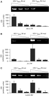
DNA integration of an HIV-1 viral clone carrying the IN K(264,266,273)R mutation is impaired. Serial dilutions of DNA extracted from CEM cells following infection with HIV wt or HIV mut were amplified with primers specific for (A) the Alu-LTR DNA fragment (to analyze the fraction of integrated HIV DNA), (B) the 2-LTR circles (to assess the levels of unintegrated HIV DNA), and (C) the lamin B2 cellular gene (to standardize the total amount of extracted DNA). The graphs were obtained by densitometric analysis of the PCR products obtained from 3–5 independent experiments.
Figure 8
Figure 8
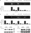
Inactivation of p300 catalytic activity inhibits HIV-1 integration. Serial dilutions of DNA extracted from SupT1 cells treated as indicated were amplified with primers specific for (A) the Alu-LTR DNA fragment and (B) the lamin B2 cellular gene. The graphs in (A) and (B) were obtained by densitometric analysis of the PCR products obtained from three independent experiments; the means±s.d. are shown. (C) Left panels: HAT assay performed on histones using immunoprecipitates obtained by incubating antibody anti-p300 or anti-GCN5 with cell extracts of SupT1 treated with SPC alone or together with Lys-CoA (upper panel: gels exposed to Cyclone screen; lower panel: Coomassie-stained gels). Right panels: SupT1 cell extracts were analyzed by immunoblot with anti-p300 or anti-GCN5 antibody (upper panels), the same blot was then incubated with anti-tubulin antibody (lower panel).

Similar articles

Cited by

References

    1. Bandyopadhyay D, Okan NA, Bales E, Nascimento L, Cole PA, Medrano EE (2002) Down-regulation of p300/CBP histone acetyltransferase activates a senescence checkpoint in human melanocytes. Cancer Res 62: 6231–6239 - PubMed
    1. Bushman FD, Engelman A, Palmer I, Wingfield P, Craigie R (1993) Domains of the integrase protein of human immunodeficiency virus type 1 responsible for polynucleotidyl transfer and zinc binding. Proc Natl Acad Sci USA 90: 3428–3432 - PMC - PubMed
    1. Cannon PM, Byles ED, Kingsman SM, Kingsman AJ (1996) Conserved sequences in the carboxyl terminus of integrase that are essential for human immunodeficiency virus type 1 replication. J Virol 70: 651–657 - PMC - PubMed
    1. Cara A, Guarnaccia F, Reitz MS, Gallo RC, Lori F (1995) Self-limiting, cell type-dependent replication of an integrase-defective human immunodeficiency virus type 1 in human primary macrophages but not T lymphocytes. Virology 208: 242–248 - PubMed
    1. Carrozza MJ, Utley RT, Workman JL, Cote J (2003) The diverse functions of histone acetyltransferase complexes. Trends Genet 19: 321–329 - PubMed

Publication types

MeSH terms