. 2005 Sep 7;24(17):3057-69.
doi: 10.1038/sj.emboj.7600765. Epub 2005 Aug 11.

The U11/U12 snRNP 65K protein acts as a molecular bridge, binding the U12 snRNA and U11-59K protein

Affiliations

The U11/U12 snRNP 65K protein acts as a molecular bridge, binding the U12 snRNA and U11-59K protein

Heike Benecke et al. EMBO J. .

Abstract

U11 and U12 interact cooperatively with the 5' splice site and branch site of pre-mRNA as a stable preformed di-snRNP complex, thereby bridging the 5' and 3' ends of the intron within the U12-dependent prespliceosome. To identify proteins contributing to di-snRNP formation and intron bridging, we investigated protein-protein and protein-RNA interactions between components of the U11/U12 snRNP. We demonstrate that the U11/U12-65K protein possesses dual binding activity, interacting directly with U12 snRNA via its C-terminal RRM and the U11-associated 59K protein via its N-terminal half. We provide evidence that, in contrast to the previously published U12 snRNA secondary structure model, the 3' half of U12 forms an extended stem-loop with a highly conserved seven-nucleotide loop and that the latter serves as the 65K binding site. Addition of an oligonucleotide comprising the 65K binding site to an in vitro splicing reaction inhibited U12-dependent, but not U2-dependent, pre-mRNA splicing. Taken together, these data suggest that U11/U12-65K and U11-59K contribute to di-snRNP formation and intron bridging in the minor prespliceosome.


Figures

Figure 1
Figure 1
The U11/U12-65K protein is evolutionarily conserved. Sequence alignment of the H. sapiens U11/U12-65K protein (gi∣48427628) with putative orthologs from M. musculus (gi∣31981077), X. laevis (deduced from ESTs gi∣26035864 and gi∣17496030; note that the 5′ end appears to be incomplete), A. thaliana (gi∣15217461) and D. melanogaster (gi∣21626517). Residues identical in at least three proteins are highlighted in black and conserved residues (gray) are grouped as follows: (D, E), (K, R), (A, S, T, G, C), (N, Q), (Y, F, M, H), (I, L, M, V) and (P). RRMs are indicated by a bar. The conserved sequence QVLHLMN(K/R)MNL is marked by dots.
Figure 2
Figure 2
65K binds U12 snRNA via its C-terminal RRM. (A) 35S-labeled, in vitro-translated 65K was incubated with in vitro-transcribed, m7G-capped U1, U2, U11 or U12 snRNA (lanes 2–5, as indicated) or no RNA (lane 6) and co-IPs with anti-cap antibodies were performed as described in Materials and methods. Lane 1: 20% of input 65K. (B) Schematic of 65K deletion mutants used in GST pulldowns. RRMs and the proline-rich region (Pro) are shaded in gray and the QVLHLMN(K/R)MNL conserved motif in black. (C–E) GST pulldowns of in vitro-transcribed, 32P-labeled UsnRNAs (indicated above each lane) by GST-65K wild-type or deletion mutants (as indicated). (C) Lane 1: 30% of a mixture of input snRNAs. (D, E) Lanes 1–4: 30 or 10% of each input snRNA. RNAs were separated by denaturing PAGE and visualized by autoradiography.
Figure 3
Figure 3
The C-terminal RRM of 65K is homologous to the N-terminal RRM of U1-A and U2-B″. (A) Domain structure of the human 65K, U1-A and U2-B″ proteins. (B) Sequence alignment of the C-terminal RRM of 65K (nt 420–503) and the N-terminal RRMs of U1-A (nt 10–89) and U2-B″ (nt 7–86). Identical amino acids are highlighted in black and conserved residues are grouped as in Figure 1. Secondary structure elements according to the atomic structures of U1-A and U2-B″ are indicated below.
Figure 4
Figure 4
The 65K protein binds the 3′ half of the U12 snRNA. (A) Co-IPs were performed as described in the legend to Figure 2 with 35S-labeled 65K and m7G-capped U11 (lane 2), U12 (lane 3) or U12 deletion mutants 5′ half, ΔSLIV, SLIII or 3′ half (lanes 4–7) or no RNA (lane 8). (B) Structure of wild-type and truncated U12 snRNAs (according to Wassarman and Steitz, 1992). 5′ half: nt 1–104; ΔSLIV: nt 1–130; SLIII: nt 79–122; 3′ half: nt 79–150. Digestion sites generating 5′ half and ΔSLIV are indicated by an arrow and nucleotides (104–130) required for 65K binding are indicated by a line.
Figure 5
Figure 5
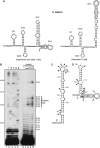
The 3′ half of U12 snRNA can adopt an alternative structure. (A) Secondary structure of the human U12 snRNA as proposed by Wassarman and Steitz (1992) (left) or generated by the MFOLD secondary structure prediction program (right). Nucleotides required for 65K binding are indicated by a line. (B) Lead(II)-induced cleavage of human U12 snRNA. Primer extension was performed after treatment of in vitro-transcribed U12 snRNA with 0, 1, 5, 10 and 50 mM lead acetate (lanes 6–10) using a primer complementary to nt 139–150. A sequencing ladder (lanes 2–5) was generated by performing primer extension of U12 snRNA in the presence of dideoxynucleotides. Lane 1: reaction without ddNTPs. Nucleotide positions are indicated on the right. Summary of cleaved sites in the alternative (C) or previously published (D) 3′ half structure of human U12 snRNA. Weak (open arrowheads) and strong (closed arrowheads) cleavage sites are indicated.
Figure 6
Figure 6
An extended 3′ SL in U12 snRNA is evolutionarily conserved. (A) Predicted secondary structures of the 3′ half of U12 from various organisms (as indicated) were generated by the MFOLD program. (B) Phylogenetic comparison of nucleotides comprising the terminal loop and adjacent stem of the 3′ SL of U12.
Figure 7
Figure 7
The 65K protein binds the terminal hairpin of the extended 3′ SL of U12. (A) Sequence of the wild-type U12 SL oligonucleotide (U12wt) and mutants (L1–L3 and S1–S5). Altered nucleotides are highlighted in black. The relative affinity of 65K-C-RRM for each mutant is indicated by plusses or a minus. (B) 65K and 65K-C-RRM interact with U12wt in GST pulldowns. (C) EMSA of 65K-C-RRM with U12wt oligo versus full-length U12 snRNA. Increasing concentrations of GST-65K-C-RRM (as indicated above each lane) were incubated with 0.01 or 0.25 pmol of the indicated oligonucleotide (lanes 1–4) or RNA (lanes 5–11), respectively. RNP complex formation was analyzed by native gel electrophoresis and visualized by autoradiography. Note that similar results were obtained when equimolar amounts (10 μM each) of U12wt oligo and U12 snRNA were used. (D) Effect of U12wt oligo mutation on 65K-C-RRM interaction. 32P-labeled oligos (as indicated above) were incubated with increasing concentrations of GST-65K-C-RRM (0, 5, 10 or 25 μM) and analyzed as in panel C.
Figure 8
Figure 8
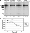
An excess of U12wt and S5 oligo inhibits P120 pre-mRNA splicing in vitro. (A) In vitro splicing was performed for 4 h with 32P-labeled P120 pre-mRNA in the presence of increasing concentrations (as indicated) of U12wt oligo (lanes 4–7), mutant L3 oligo (lanes 8–11) or mutant S5 oligo (lanes 12–15). Lanes 1–3: P120 splicing in the absence of oligo after 0, 2 and 4 h. RNA was separated on an 8% polyacrylamide–8 M urea gel and visualized by autoradiography. The positions of the splicing intermediates and products are indicated schematically on the left. (B) Quantitation of splicing efficiency. The amounts of pre-mRNA and mRNA were determined using a PhosphorImager and the ratio of mRNA to pre-mRNA was plotted as a function of oligonucleotide concentration.
Figure 9
Figure 9
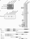
The N-terminal half of 65K interacts with the C-terminal 149 aa of the 59K protein. (A) Yeast two-hybrid assays with the 65K and 59K proteins (and deletion mutants thereof). (B) 65K and 59K interact in Far Western overlays. U11/U12 proteins on nitrocellulose strips were visualized by staining with Ponceau S (lane 1) or incubated with 35S-labeled, in vitro-translated protein (as indicated above each lane) and visualized by fluorography (lanes 2–4). U11/U12 proteins are indicated on the left. (C) GST pulldowns with 35S-labeled 59K332–481 and GST (lane 2), or GST fusions of 65K and deletion mutants thereof (lanes 3–7; as indicated). Co-precipitated protein or 10% of the input (lane 1) was analyzed by SDS–PAGE and visualized by fluorography. (D) Schematic of wild-type and truncated 65K and 59K proteins used in panels A–C. Abbreviations are as in Figure 2. Arginine- (Arg) and glutamic acid-rich regions (Glu) are shaded gray.

References

    1. Achsel T, Ahrens K, Brahms H, Teigelkamp S, Lührmann R (1998) The human U5-220kD protein (hPrp8) forms a stable RNA-free complex with several U5-specific proteins, including an RNA unwindase, a homologue of ribosomal elongation factor EF-2, and a novel WD-40 protein. Mol Cell Biol 18: 6756–6766 - PMC - PubMed
    1. Bandziulis RJ, Swanson MS, Dreyfuss G (1989) RNA-binding proteins as developmental regulators. Genes Dev 3: 431–437 - PubMed
    1. Bentley RC, Keene JD (1991) Recognition of U1 and U2 small nuclear RNAs can be altered by a 5-amino-acid segment in the U2 small nuclear ribonucleoprotein particle (snRNP) B′′ protein and through interactions with U2 snRNP-A′ protein. Mol Cell Biol 11: 1829–1839 - PMC - PubMed
    1. Birney E, Kumar S, Krainer AR (1993) Analysis of the RNA-recognition motif and RS and RGG domains: conservation in metazoan pre-mRNA splicing factors. Nucleic Acids Res 21: 5803–5816 - PMC - PubMed
    1. Brunel C, Romby P (2000) Probing RNA structure and RNA–ligand complexes with chemical probes. Methods Enzymol 318: 3–21 - PubMed

Publication types

Substances

LinkOut - more resources