

Microcolumn separation of amine metabolites in the fruit fly
- PMID: 16097779
- PMCID: PMC1351352
- DOI: 10.1021/ac050474m
Microcolumn separation of amine metabolites in the fruit fly
Abstract
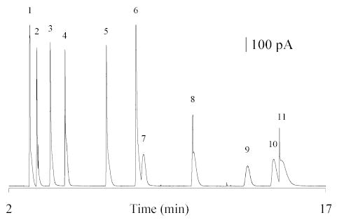
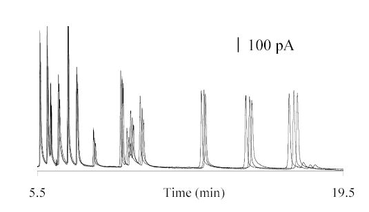
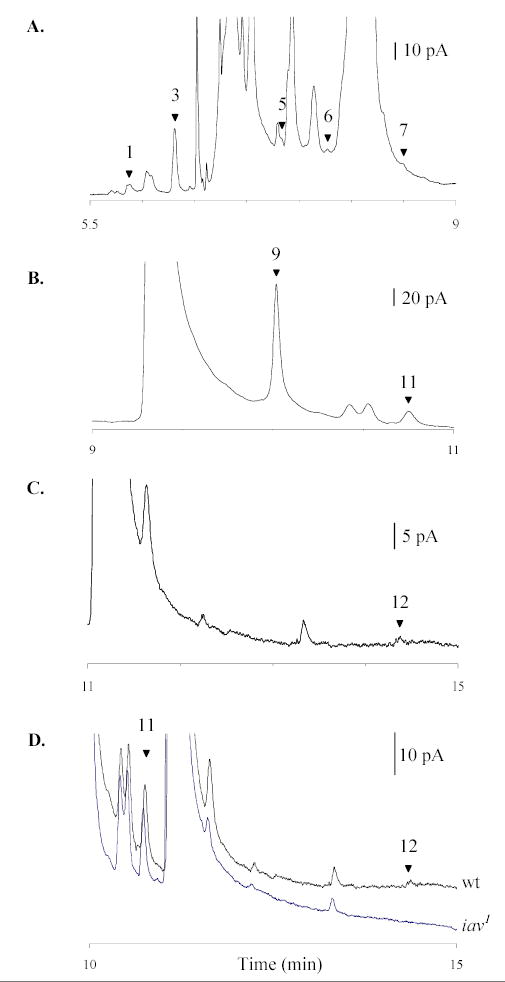
Electrophoretic resolution of 14 biogenic amines and metabolites with similar mobilities is addressed by employing micellar electrokinetic capillary chromatography coupled to amperometric electrochemical detection. The present study describes the optimization of separation conditions to achieve resolution of analytes of biological significance within 20 min in a single separation. They include dopamine, epinephrine, norepinephrine, octopamine (OA), L-3, 4-dihydroxyphenylalanine, tyramine (TA), and serotonin as well as metabolites 5-hydroxyindolacetic acid, 3,4-dihydroxyphenylacetic acid, homovanillic acid, and 3-methoxytyramine in addition to N-acetylated metabolites including N-acetyldopamine, N-acetyloctopamine (naOA), and N-acetylserotonin. The optimized conditions used result in excellent reproducibility and predictable peak shifting, thus enabling identification of several metabolites along with their biogenic amine precursors in biological samples, specifically from the fruit fly Drosophila melanogaster. The separation method is sensitive, selective, and quantitative as demonstrated by its capacity to detect changes in TA, OA, and naOA present in the head homogenates of the Canton-S and mutant inactive(1) Drosophila lines. Quantitative analysis of metabolites in conjunction with their biogenic amine precursors in a single separation offers tremendous potential to understand the physiological processes and underlying mechanisms mediated by various biogenic amines in Drosophila and other animals.
Figures

References
-
- Peaston RT, Weinkove C. Ann Clin Biochem. 2004;41:17–38. - PubMed
-
- Terabe S, Markuszewski MJ, Inoue N, Otsuka K, Nishioka T. Pure Appl Chem. 2001;73:1563–1572.
-
- Wallingford RA, Ewing AG. Anal Chem. 1988;60:258–263. - PubMed
-
- Wallingford RA, Ewing AG. J Chromatogr. 1988;441:299–309. - PubMed
-
- Wallingford RA, Curry PD, Ewing AG. J Microcolumn Sep. 1989;1:23–27.
Publication types
MeSH terms
Substances
Grants and funding
LinkOut - more resources
Full Text Sources
Molecular Biology Databases
