Comparative Study
. 2005 Dec 1;106(12):3803-10.
doi: 10.1182/blood-2005-01-0179. Epub 2005 Aug 11.

Analysis of thrombocyte development in CD41-GFP transgenic zebrafish

Affiliations
Comparative Study

Analysis of thrombocyte development in CD41-GFP transgenic zebrafish

Hui-Feng Lin et al. Blood. .

Abstract

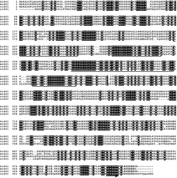
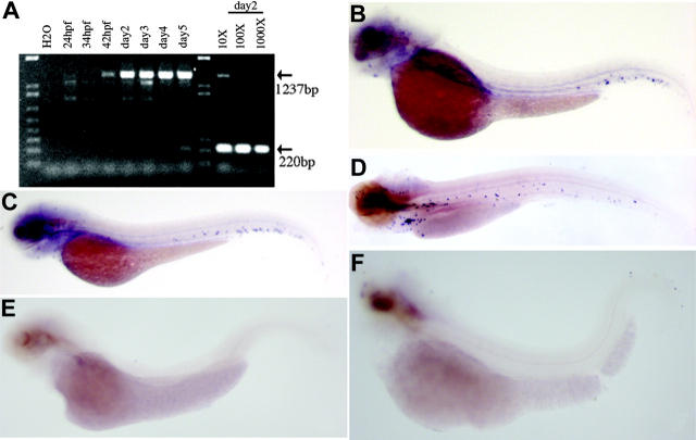
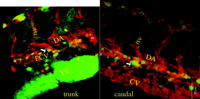
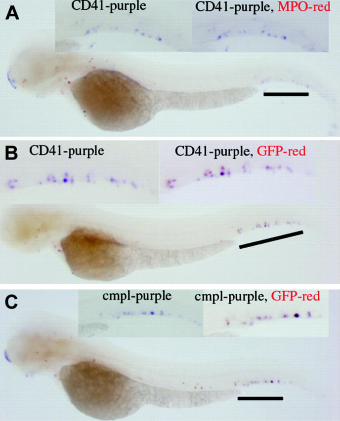
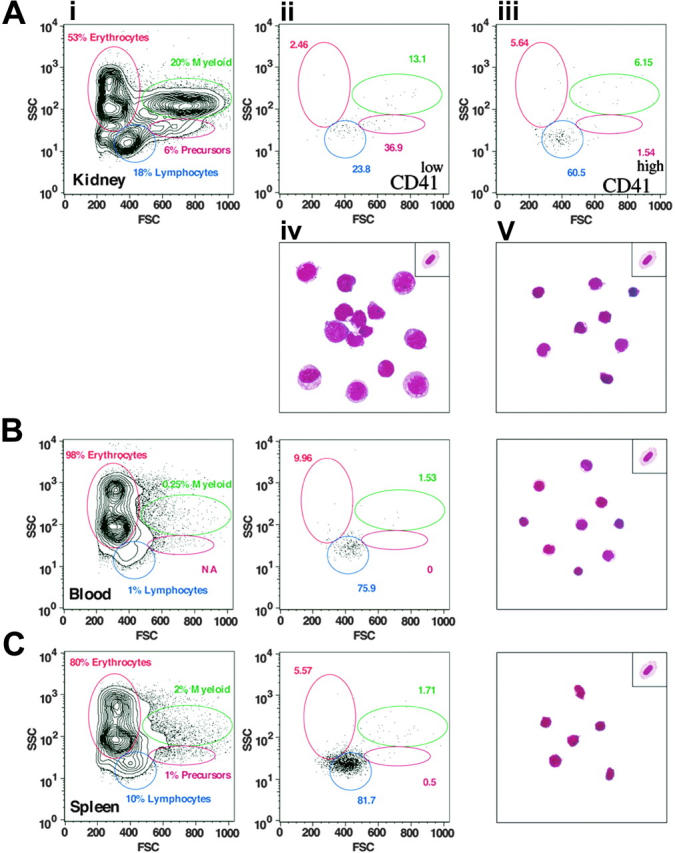
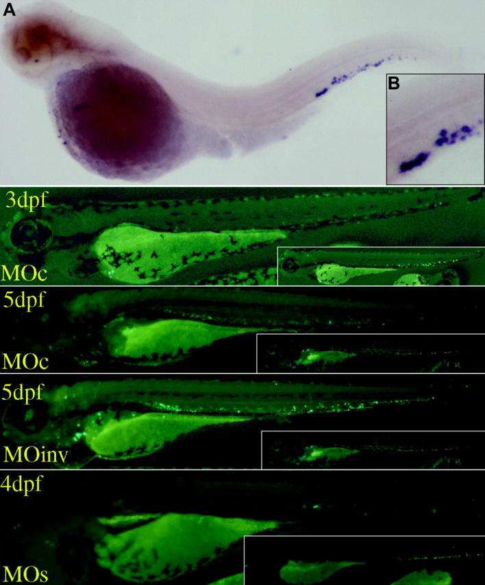
Thrombocytes are the nucleated equivalent of platelets in nonmammalian vertebrates such as the zebrafish, Danio rerio. We have cloned zebrafish CD41 cDNA (alpha(IIb), glycoprotein IIb [GPIIb]) and its promoter and have generated transgenic zebrafish lines with green fluorescent protein (GFP)-tagged thrombocytes. CD41 mRNA transcripts appeared 42 hours after fertilization (hpf) by reverse-transcriptase-polymerase chain reaction (RT-PCR) and at 48 hpf in circulating hematopoietic cells. Flow sorting of thrombocytes from the mesonephros of adult CD41-GFP zebrafish showed a GFP(high) subset, which had the morphologic appearance of mature thrombocytes, and a GFP(low) subset with an immature appearance, suggesting that they may be thrombocyte precursors. Confocal laser microscopy of embryos 40 and 48 hpf also showed a nonmobile population of GFP+ cells in a discrete area between the dorsal aorta and caudal vein. Production of circulating thrombocytes was inhibited by the injection of antisense morpholinos for the stem-cell transcription factor scl and c-mpl, the receptor for thrombopoietin. The nonmobile pool of GFP+ cells was abolished by scl knockdown and partially inhibited by c-mpl knockdown. These studies have shown that it is possible to identify thrombocytes, thrombocyte precursors, and, possibly, early hematopoietic stem cells in zebrafish embryos and track their proliferation and maturation.


Figures

Figure 1.
Figure 1.
Alignment of deduced CD41 protein sequence. Alignment of the deduced amino acid sequences of the human (hcd41), mouse (mcd41), rat (rcd41), and zebrafish (zcd41) CD41 is shown. Forty percent of the zebrafish sequence is identical to the sequences of the other three species. There are some conserved features, including the location of cysteine residues, the four potential calcium-binding domains (underlined), and the C-terminal GFFKR sequence (underlined with arrow). The regions of sequence identity in the four species are shaded.
Figure 2.
Figure 2.
Results of RT-PCR and whole-mount in situ hybridization of CD41. (A) Shown is the analysis of the expression of CD41 mRNA in developing zebrafish embryos by RT-PCR. The CD41 transcript is first detected at 42 hpf. Dilutions (10 ×, 100 ×, and 1000 ×) of the cDNA preparation 2 dpf, with EF-1α as a control, indicated that CD41 transcript is at least 1000 × lower than EF-1α transcript. The expected PCR product for CD41 is 1237 bp and for EF-1α is 220 bp. (B-D) Shown also are results of whole-mount in situ hybridization of wild-type zebrafish embryos with zebrafish CD41 antisense cRNA. (B) In embryos 2 dpf, punctate purple CD41+ cells are present in the ventral region of the dorsal aorta. (C) In embryos 3 dpf, the number of CD41+ cells has increased and they appear in the circulation. (D) In embryos 5 dpf, more CD41+ cells are present in the circulation. The CD41 transcript is not detected in the spadetail (E) and cloche (F) embryos 3 dpf.
Figure 3.
Figure 3.
Expression of GFP in the CD41-GFP embryos. Photomicrographs showing CD41-GFP+ expression in transgenic embryos are presented. Unfertilized eggs from homozygous (A) and wild-type (B) females demonstrate the maternally derived GFP. At 48 hpf, bright CD41-GFP+ cells are detected in the region between dorsal aorta and caudal vein (arrows, C) and cardiac sinus/yolk sac (arrow near yolk). By 3 dpf, increasing numbers of CD41-GFP+ cells are detected in a similar region (arrows near tail, D), and the cardiac sinus/yolk sac (arrow near yolk), as well as in the circulation (D). By 5 dpf, the majority of the CD41-GFP+ cells are circulating, and a collection of CD41-GFP+ cells appears near the developing mesonephros (arrow, E).
Figure 4.
Figure 4.
Confocal scanning microscopy of CD41-GFP/LMO2-dsRed embryos. Shown are 2-dimensional photomicrographs of confocal microscopy near the trunk and caudal portion of an embryo 48 hpf. Two corresponding movies of 3-dimensional reconstructions of the same embryo are available as supplemental material on the Blood website; click on the Supplemental Movies link at the top of the online article. Of note is that the major vessels, whose endothelial cells are tagged with LMO2-dsRed, emit red fluorescence, and the nonmobile CD41-GFP+ cells emit green fluorescence. The GFP+ cells are extravascular and are clearly located in the region between the dorsal aorta and the posterior cardinal vein (trunk view, arrow) and caudal portion between the caudal dorsal aorta and caudal vein (caudal view, arrow). DA indicates dorsal aorta; PCV, posterior cardinal vein; ISV, intersomitic vessel; YE, yolk extension; and CV, caudal vein.
Figure 5.
Figure 5.
Two-color whole-mount in situ hybridization. (A) CD41 expression does not colocalize with MPO, as judged by the CD41 cells (purple) that do not turn red in the in situ result. (B) On the contrary, the coexpression of CD41 and GFP is supported by the red color (GFP) outcome of originally purple cells (CD41). (C) In addition, coexpression of c-mpl and GFP in the same cells is supported.
Figure 6.
Figure 6.
Results of FACS analyses. (A) Panel i shows 2 populations of GFP+ cells (CD41low and CD41high representing 0.81% and 0.79% of WKM, respectively) that are detected in the kidney. CD41low cells (ii) are predominantly large prothrombocytic cells and CD41high cells (iii) are differentiated thrombocytes as demonstrated by the corresponding Wright-Giemsa staining of flow-sorted cells shown in panels iv and v (see “Flow sorting of GFP+ thrombocytes”). A representative erythrocyte is placed at the top right corner for comparison. By contrast, the vast majority of cells isolated from peripheral blood (B) and spleen (C) are CD41high (0.64% and 4.21%, respectively) and display the mature thrombocyte morphology.
Figure 7.
Figure 7.
Results of whole-mount in situ hybridization of c-mpl and c-mpl and scl morpholino injection. Shown are the results of whole-mount in situ hybridization with antisense c-mpl cRNA (A; high power, B) at the embryo 3 dpf. c-mpl+ cells are detected in the same location as CD41+ and GFP+ cells detected in previous studies. Injection of a zebrafish c-mpl antisense morpholino (MOc) reduced the number of GFP+ cells 3 and 5 dpf. c-mpl morphants have some residual GFP+ cells. Injection of an inverted morpholino (MOinv) did not decrease the number of circulating GFP+ cells. Injection of 2 scl morpholinos (MOs) also reduced the number of GFP+ cells in the morphants at 4 dpf, in addition to causing tail deformation. For comparison, uninjected siblings of the same developmental stages are posted in the right corner of each picture.

References

    1. Jagadeeswaran P, Sheehan JP, Craig FE, Troyer D. Identification and characterization of zebrafish thrombocytes. Br J Haematol. 1999;107: 731-738. - PubMed
    1. Hartwig J, Italiano J Jr. The birth of the platelet. J Thromb Haemost. 2003;1: 1580-1586. - PubMed
    1. Nakorn TN, Miyamoto T, Weissman IL. Characterization of mouse clonogenic megakaryocyte progenitors. Proc Natl Acad Sci U S A. 2003;100: 205-210. - PMC - PubMed
    1. McEver RP, Beckstead JH, Moore KL, Marshall-Carlson L, Bainton DF. GMP-140, a platelet alpha-granule membrane protein, is also synthesized by vascular endothelial cells and is localized in Weibel-Palade bodies. J Clin Invest. 1989;84: 92-99. - PMC - PubMed
    1. Poncz M, Surrey S, LaRocco P, et al. Cloning and characterization of platelet factor 4 cDNA derived from a human erythroleukemic cell line. Blood. 1987;69: 219-223. - PubMed

Publication types

Substances