. 2005 Nov;89(5):3337-44.
doi: 10.1529/biophysj.105.062034. Epub 2005 Aug 12.

Ligand binding modulates the mechanical stability of dihydrofolate reductase

Affiliations

Ligand binding modulates the mechanical stability of dihydrofolate reductase

Sri Rama Koti Ainavarapu et al. Biophys J. 2005 Nov.

Abstract

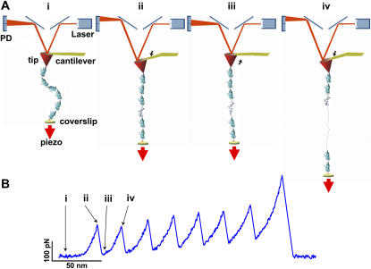
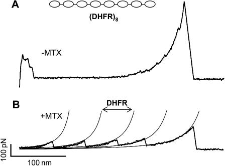
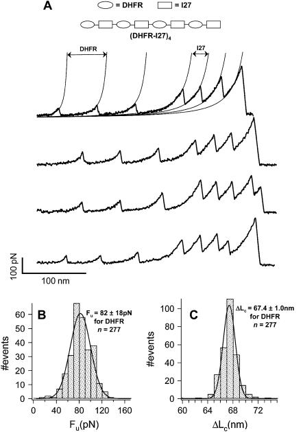
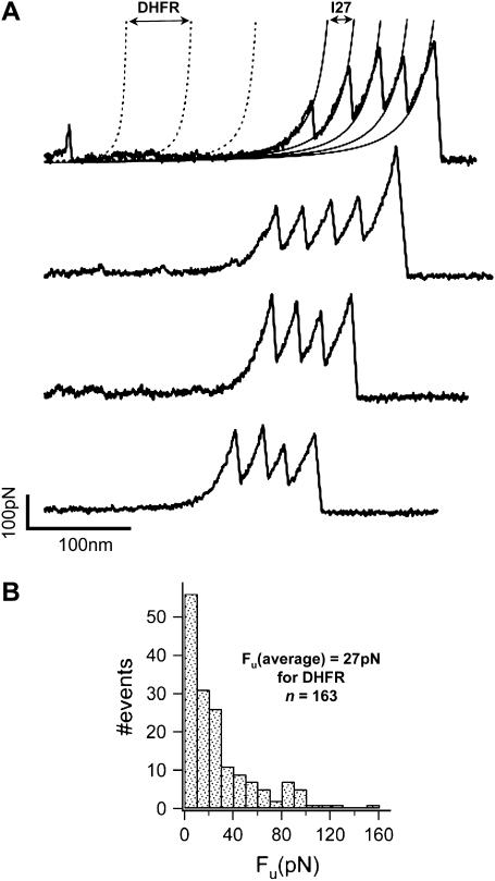
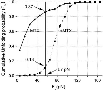
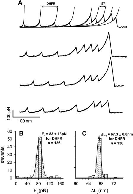
We use single-molecule force spectroscopy to demonstrate that the mechanical stability of the enzyme dihydrofolate reductase (DHFR) is modulated by ligand binding. In the absence of bound ligands, DHFR extends at very low forces, averaging 27 pN, without any characteristic mechanical fingerprint. By contrast, in the presence of micromolar concentrations of the ligands methotrexate, nicotinamide adenine dihydrogen phosphate, or dihydrofolate, much higher forces are required (82 +/- 18 pN, 98 +/- 15 pN, and 83 +/- 16 pN, respectively) and a characteristic fingerprint is observed in the force-extension curves. The increased mechanical stability triggered by these ligands is not additive. Our results explain the large reduction in the degradation rate of DHFR, in the presence of its ligands. Our observations support the view that the rate-limiting step in protein degradation by adenosine triphosphate-dependent proteases is the mechanical unfolding of the target protein.


Figures

FIGURE 1
FIGURE 1
Mechanical stretching of a polyprotein using single-molecule atomic force microscopy. (A) (i) A single polyprotein molecule is held between the cantilever tip and the coverslip, whose position can be controlled with high precision using a piezoelectric positioner (piezo). (ii) Moving the coverslip away from the tip exerts a stretching force on the polyprotein, which in turn bends the cantilever. The bending of the cantilever changes the position of the laser beam on the split photo diode (PD), registering the pulling force. The applied force can be determined from the spring constant of the cantilever and the degree of cantilever bending. At this high pulling force, a protein domain unfolds. (iii) The unfolded domain can now readily extend, relaxing the cantilever. (iv) The piezo continues to move, stretching the polyprotein to a new high force peak, repeating the sequence until the whole polyprotein has unfolded. This process results in a force-extension curve with a characteristic sawtooth pattern shape. (B) A typical sawtooth pattern curve obtained by stretching an I278 polyprotein (28). The labels iiv represent the sequence of events shown in A.
FIGURE 2
FIGURE 2
The force necessary to unfold DHFR is dependent on the presence of the ligand Methotrexate (MTX). (A) Force-extension curve obtained by stretching the polyprotein DHFR8 in the absence of methotrexate (MTX). The lack of a sawtooth pattern suggests that DHFR is mechanically weak. However, absence of a fingerprint makes it difficult to be certain that such recordings correspond to DHFR and not from a contaminating molecule. (B) Force-extension curve obtained by stretching the polyprotein DHFR8 in the presence of 1.2 mM MTX. We now observe a force-extension curve showing a clear sawtooth pattern of unfolding events at an average force of 78 pN ± 14 (n = 72; see Table 1). Fits of the wormlike chain (WLC) model of polymer elasticity (thin lines) reveal a contour increment between unfolding events of ΔLc = 67.3 ± 0.5 nm (n = 72), which is in close agreement with the expected length gained by unfolding a DHFR molecule (65 nm).
FIGURE 3
FIGURE 3
Use of a polyprotein chimera to probe the mechanical stability of DHFR. (A) A panel of four force-extension curves obtained by stretching the (DHFR-I27)4 polyprotein chimera, in the presence of 190 μM MTX. The low unfolding force and much larger contour length easily distinguish the DHFR unfolding events (first) from the I27 unfolding events (last). Fits of the WLC model (thin lines) to the data are used to measure the contour length increment between unfolding events. (B) Histogram of unfolding forces for the DHFR-MTX complex. A Gaussian fit (thin line) gives 82 ± 18 pN (n = 277). (C) Histogram of contour length increments measured with the WLC. A Gaussian fit (thin line) gives 67.4 ± 1.0 nm (n = 277). By contrast, the unfolding force measured from the I27 peaks is 220 ± 36 pN and the contour length increment is 28.0 ± 0.7 nm (n = 322).
FIGURE 4
FIGURE 4
The mechanical stability of DHFR can be unambiguously determined using the (DHFR-I27)4 polyprotein chimera. (A) A panel of four force-extension curves obtained by stretching (DHFR-I27)4 in the absence of added MTX. The principal feature of these recordings is the long featureless spacer that we observe preceding the I27 unfolding events. The long spacer is marked occasionally by some low level unfolding peaks. By contrast, the I27 unfolding events can be readily observed and quantified with fits of the WLC (thin lines). The unfolding force of I27 is 209 ± 34 pN and the increment of contour length upon I27 unfolding is 28.1 ± 1.3 nm (n = 226). To estimate the force required to unfold DHFR, we generate a series of WLC curves, equally spaced by 67 nm (dotted lines), starting backward from the first I27 unfolding event. The intersection between these WLC curves and the experimental values are taken as the unfolding force for DHFR. (B) Histogram of the unfolding forces of DHFR. The mean unfolding force for DHFR is 27 pN (n = 163).
FIGURE 5
FIGURE 5
Plot of the cumulative unfolding probability as a function of unfolding force in the presence and absence of MTX at a constant pulling rate of 400 nm/s and a constant unfolding rate of ∼6/s. The cumulative probability of unfolding was calculated by integrating the histograms of Fig. 3 B (+MTX) and Fig. 4 B (−MTX), and then normalizing them to 1. The thick vertical line marks a pulling force of 57 pN (see text). At this pulling force DHFR is most likely to be unfolded (Pu = 0.87). By contrast, the DHFR-MTX complex is most likely to remain folded (Pu = 0.13).
FIGURE 6
FIGURE 6
The mechanical stability of DHFR-MTX complex is not increased by adding a second ligand. (A) A panel of force-extension curves measured by stretching the (DHFR-I27)4 polyprotein chimera in the presence of 190 μM MTX and 210 μM NADPH. Despite the fact that both MTX and NADPH stabilize DHFR to a similar extent (see Table 2), their stabilizing effects are not additive. As before, we measure the magnitude of the unfolding peaks for both DHFR and the I27 fingerprint (see Table 2). The thin lines are WLC fits. (B) Histogram of unfolding forces of DHFR in the presence of MTX and NADPH. A Gaussian fit (thin line) gives an unfolding force of 83 ± 13 pN (n = 136). (C) Histogram of contour length increments after DHFR unfolding measured with the WLC. A Gaussian fit (thin line) gives 67.3 ± 0.8 nm (n = 136). These results are indistinguishable from those obtained with either MTX or NADPH alone, indicating that their effect on the mechanical stability of DHFR is not additive.

Comment in

  • Fingerprinting DHFR in single-molecule AFM studies.
    Ainavarapu SR, Li L, Fernandez JM. Ainavarapu SR, et al. Biophys J. 2006 Sep 1;91(5):2009-10, discussion 2011-2. doi: 10.1529/biophysj.106.085126. Epub 2006 Jun 16. Biophys J. 2006. PMID: 16782796 Free PMC article. No abstract available.

Similar articles

Cited by

References

    1. Schnell, J. R., H. J. Dyson, and P. E. Wright. 2004. Structure, dynamics, and catalytic function of dihydrofolate reductase. Annu. Rev. Biophys. Biomol. Struct. 33:119–140. - PubMed
    1. Benkovic, S. J., and S. Hammes-Schiffer. 2003. A perspective on enzyme catalysis. Science. 301:1196–1202. - PubMed
    1. Pineda, P., A. Kanter, R. S. McIvor, S. J. Benkovic, A. Rosowsky, and C. R. Wagner. 2003. Dihydrofolate reductase mutant with exceptional resistance to methotrexate but not to trimetrexate. J. Med. Chem. 46:2816–2818. - PubMed
    1. Davies, J. F., T. J. Delcamp, N. J. Prendergast, V. A. Ashford, J. H. Freisheim, and J. Kraut. 1990. Crystal structures of recombinant human dihydrofolate reductase complexed with folate and 5-deazafolate. Biochemistry. 29:9467–9479. - PubMed
    1. Focia, P. J., I. V. Shepotinovskaya, J. A. Seidler, and D. M. Freymann. 2004. Heterodimeric GTPase core of the SRP targeting complex. Science. 303:373–377. - PMC - PubMed

Publication types

MeSH terms

LinkOut - more resources