. 2005 Nov;89(5):3223-34.
doi: 10.1529/biophysj.104.057505. Epub 2005 Aug 12.

The E-hook of tubulin interacts with kinesin's head to increase processivity and speed

Affiliations

The E-hook of tubulin interacts with kinesin's head to increase processivity and speed

Stefan Lakämper et al. Biophys J. 2005 Nov.

Abstract

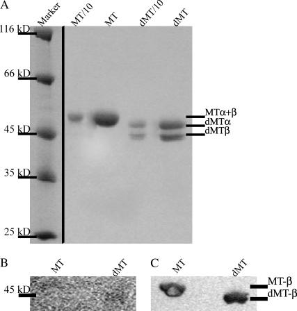
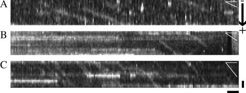
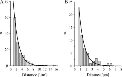
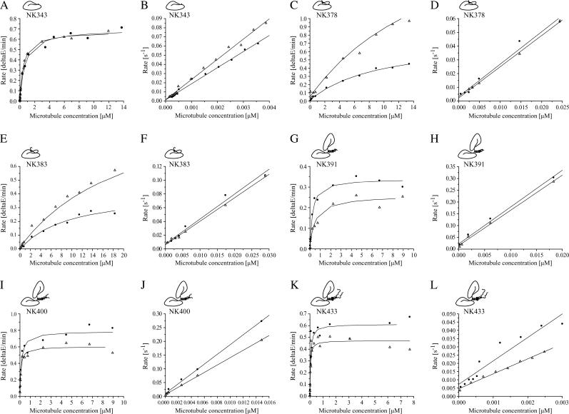
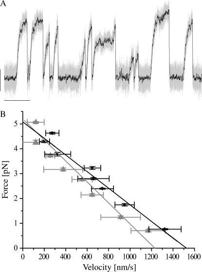
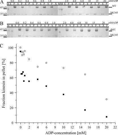
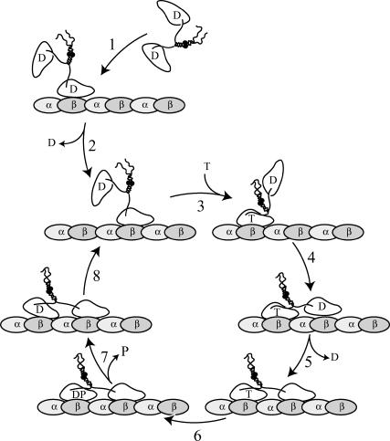
Kinesins are dimeric motor proteins that move processively along microtubules. It has been proposed that the processivity of conventional kinesins is increased by electrostatic interactions between the positively charged neck of the motor and the negatively charged C-terminus of tubulin (E-hook). In this report we challenge this anchoring hypothesis by studying the motility of a fast fungal kinesin from Neurospora crassa (NcKin). NcKin is highly processive despite lacking the positive charges in the neck. We present a detailed analysis of how proteolytic removal of the E-hook affects truncated monomeric and dimeric constructs of NcKin. Upon digestion we observe a strong reduction of the processivity and speed of dimeric motor constructs. Monomeric motors with truncated or no neck display the same reduction of microtubule gliding speed as dimeric constructs, suggesting that the E-hook interacts with the head only. The E-hook has no effect on the strongly bound states of NcKin as microtubule digestion does not alter the stall forces produced by single dimeric motors, suggesting that the E-hook affects the interaction site of the kinesin.ADP-head and the microtubule. In fact, kinetic and binding experiments indicate that removal of the E-hook shifts the binding equilibrium of the weakly attached kinesin.ADP-head toward a more strongly bound state, which may explain reduced processivity and speed on digested microtubules.


Figures

FIGURE 1
FIGURE 1
MT digestion. Panel A shows an SDS-PAGE analysis of native MTs and subtilisin dMTs. Undigested α- and β-subunits of tubulin do not separate under standard conditions. Digestion, however, leads to clear separation of the subunits. We confirmed complete digestion of β-tubulin using a Western blot with monoclonal anti-β-tubulin antibodies. Panel B shows the Ponceau S-stained blot, and panel C the Western blot after detection of the primary antibody with a secondary, alkaline phosphatase-conjugated antibody and incubation with NBT/BCIP as substrate.
FIGURE 2
FIGURE 2
Kymographs of single molecule fluorescence events. Kymographs were generated from unprocessed image sequences using Metamorph software. From each frame a single row of pixels along the length of the MT was extracted. The resulting slices from sequential frames were aligned side-by-side such that the vertical axis of the kymographs represents the direction along the MT (scale bar = 1 μm) and the horizontal axis represents the time (scale bar corresponds to 1 s). Events of single fluorescently labeled motor molecules moving along an MT emerge as inclined bright lines, and immotile motors or fluorescent particles lead to horizontal streaks. In this panel, all MTs are oriented with the plus end of the MT pointing downward. Panels A and C show examples of HsKin560 and NK433 motors, respectively, moving along an MT. Panel B shows examples of single NK433 motors moving along a dMT. White angles in the far upper corner of each panel illustrate the different slopes of the events, reflecting the speed of the motors.
FIGURE 3
FIGURE 3
Histogram analysis of single molecule fluorescence assays. The distances traveled in individual events of processive movement along the MTs were combined in a histogram. The data were fit by a single exponential, the time constant of which represents the average run length. Panel A shows data for NcKin on MTs (data from Lakamper et al. (24); N = 182; run length 1.75 ± 0.09 μm). Panel B shows the histogram for NcKin motors moving along subtilisin-treated MTs (N = 67; run length 0.89 ± 0.08 μm, Table 1).
FIGURE 4
FIGURE 4
Kinetic analysis of NcKin motors interacting with MTs and dMTs. Panels AL show typical ATPase and ADP-release diagrams of MTs (circles) and dMTs (triangles) for the motor constructs used in this study ordered in increasing length. MT digestion leaves the kcat of the minimal motor domain, NK343, unaffected (A; MT 247.6 ± 22.2 ATP/head*s; dMT 249.4 ± 17.0 ATP/head*s). The slight reduction of the k0.5,MT-value upon digestion of MTs from 0.94 ± 0.35 μM to 0.76 ± 0.21 μM is statistically not significant. (B) The rates of mantADP release for NK343 increase from 19.6 ± 0.94 μM−1s−1 on MTs to 24.75 ± 2.78 μM−1s−1. ATP turnover of NK378 on dMTs (C) is increased from 18.81 ± 1.69 ATP/head*s to 33.64 ± 2.89 ATP/head*s, whereas k0.5,MT is doubled from 7.44 ± 1.21 μM to 13.13 ± 1.54 μM on dMTs. Kbi,ADP-values drop from 2.58 ± 0.30 μM−1s−1 on MTs to 2.05 ± 0.08 μM−1s−1 on dMTs (D). Similarly, ATP turnover of NK383 on dMTs (E) is increased from 20.53 ± 2.71 ATP/head*s to 32.92 ± 5.56 ATP/head*s, whereas k0.5,MT is doubled from 6.76 ± 1.50 μM to 11.72 ± 3.14 μM on dMTs. kbi,ADP-values drop from 3.44 ± 0.13μM−1s−1 on MTs to 3.18 ± 0.13 μM−1s−1 on dMTs (F). ATP turnover of dimeric NK391 on dMTs (G) is reduced from 71.72 ± 5.16 ATP/head*s to 50.01 ± 4.10 ATP/head*s, whereas k0.5,MT is increased from 0.21 ± 0.05 μM to 0.31 ± 0.08 μM on dMT. kbi,ADP-values remain unchanged at 14.16 ± 1.02 μM−1s−1 and 14.99 ± 0.72 μM−1s−1 (H). ATP turnover of NK400 on dMTs similarly drops from 68.59 ± 7.35 ATP/head*s to 49.84 ± 6.00 ATP/head*s, k0.5,MT increases from 0.11 ± 0.01 μM to 0.16 ± 0.05 μM on dMTs (I), whereas kbi,ADP-values drop from 20.44 ± 1.55 μM−1s−1 on MTs to 14.12 ± 0.65 μM−1s−1 on dMTs (J). ATP turnover of NK433 on dMTs (K) is decreased from 64.18 ± 4.45 ATP/head*s to 46.10 ± 4.16 ATP/head*s, whereas k0.5,MT is doubled from 0.23 ± 0.06 μM to 0.46 ± 0.13 μM on dMTs. kbi,ADP values for NK433 drop from 16.57 ± 1.47 μM−1s−1 to 9.77 ± 0.99 μM−1s−1 (L) upon digestion.
FIGURE 5
FIGURE 5
Single molecule trapping experiments. Panel A shows a representative tracing of a trapped, kinesin-coated bead powered by a single NK433 dimer along a dMT (unfiltered position signal, light shaded; filtered at 50 Hz, solid; vertical scale 50 nm, horizontal scale 2 s, trap stiffness 0.049 pN/nm). Using custom-written C-code, we fit a series of short linear regressions (20 nm displacement, duration >20 ms) to individual runs of kinesin and computed the average speed and force for each segment. Speeds were averaged over 0.5 pN force intervals and combined to a force-velocity diagram. Panel B shows examples of force-velocity diagrams for single NK433 motors interacting with MTs (solid) and dMTs (shaded) at 2 mM ATP. These two force-velocity relations were averaged from traces containing a large number of individual events all obtained from a single kinesin-coated bead. The extrapolated initial velocity at zero-load conditions for dMTs is ∼20% lower (1.52 ± 0.10 μm/s) than for MTs (1.22 ± 0.12 μm/s). At higher forces, the two curves converge to indistinguishable stall forces of 5.08 ± 0.20 pN and 5.20 ± 0.23 pN for MTs and dMTs, respectively (the data from five such measurements are summarized in Table 4).
FIGURE 6
FIGURE 6
Equilibrium-binding experiments with NK343. Panels A and B show the results from binding experiments of NK343 to MTs and dMTs, respectively (M = marker, S = supernatant, P = pellet). Panel C illustrates the fraction of motor bound to the MTs (▪) and dMTs (○) as a function of the ADP concentration. Notice that much larger fractions of motor partition to the dMTs at any ADP concentration.
FIGURE 7
FIGURE 7
Schematic working hypothesis for the processive movement of NcKin. Proposed influence of the E-hook on the chemomechanical cycle of kinesin. See Discussion for a detailed description of the model. In the native MT-motor system, the E-hook modulates (reduces) the affinity of the kinesin·ADP complex such that weak-binding intermediates facilitate fast, processive movement of kinesin. Digestion of the MT (removal of the E-hook) creates a trapped ADP-state(s) of kinesin: we propose that the forward rates of steps 5 and/or 8 in our model are severely slowed in the absence of the E-hook, leading to the observed changes in gliding speed and processivity.

Similar articles

Cited by

References

    1. Vale, R. D. 2003. The molecular motor toolbox for intracellular transport. Cell. 112:467–480. - PubMed
    1. Vale, R. D., T. S. Reese, and M. P. Sheetz. 1985. Identification of a novel force-generating protein, kinesin, involved in microtubule-based motility. Cell. 42:39–50. - PMC - PubMed
    1. Brady, S. T. 1985. A novel brain ATPase with properties expected for the fast axonal transport motor. Nature. 317:73–75. - PubMed
    1. Kuznetsov, S. A., E. A. Vaisberg, N. A. Shanina, N. N. Magretova, V. Y. Chernyak, and V. I. Gelfand. 1988. The quaternary structure of bovine brain kinesin. EMBO J. 7:353–356. - PMC - PubMed
    1. Scholey, J. M., J. Heuser, J. T. Yang, and L. S. Goldstein. 1989. Identification of globular mechanochemical heads of kinesin. Nature. 338:355–357. - PubMed

Publication types

MeSH terms

LinkOut - more resources