

doi: 10.1038/ncb1289.
Epub 2005 Aug 14.
Protein kinase D regulates vesicular transport by phosphorylating and activating phosphatidylinositol-4 kinase IIIbeta at the Golgi complex
Affiliations
- PMID: 16100512
- PMCID: PMC1458033
- DOI: 10.1038/ncb1289
Item in Clipboard
Protein kinase D regulates vesicular transport by phosphorylating and activating phosphatidylinositol-4 kinase IIIbeta at the Golgi complex
Nat Cell Biol.
2005 Sep.
Abstract
Protein kinase D (PKD) regulates the fission of vesicles originating from the trans-Golgi network. We show that phosphatidylinositol 4-kinase IIIbeta (PI4KIIIbeta) - a key player in the structure and function of the Golgi complex - is a physiological substrate of PKD. Of the three PKD isoforms, only PKD1 and PKD2 phosphorylated PI4KIIIbeta at a motif that is highly conserved from yeast to humans. PKD-mediated phosphorylation stimulated lipid kinase activity of PI4KIIIbeta and enhanced vesicular stomatitis virus G-protein transport to the plasma membrane. The identification of PI4KIIIbeta as one of the PKD substrates should help to reveal the molecular events that enable transport-carrier formation.
Figures
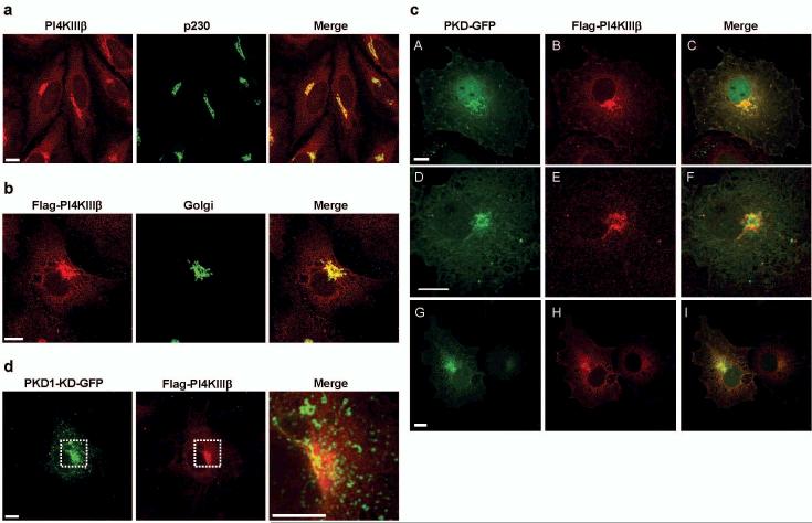
PKD-GFP and Flag-PI4KIIIβ colocalize at the Golgi compartment. (a) PI4KIIIβ localizes to the Golgi complex. Hela cells were double-labelled with antibodies against PI4KIIIβ (red) and a trans-Golgi protein p230 (green). (b) Flag-PI4KIIIβ localizes to the Golgi complex. COS7 cells expressing Flag-PI4KIIIβ and a Golgi-specific YFP tagged protein (green) were stained with antibodies against PI4KIIIβ (red). (c) COS7 cells expressing wildtype PKD-GFP fusion proteins (green, A: PKD1-GFP; D: PKD2-GFP; G: PKD3-GFP) and Flag-PI4KIIIβ (B, E, H) were labelled with antibodies against PI4KIIIβ (red). (d) COS7 cells expressing a kinase-dead PKD1-GFP fusion protein (PKD1-KD-GFP, green) and Flag-PI4KIIIβ (red). Scale bar represents 10μm. Shown are parallel projections of stacks taken at 0.5 μm distance in z-direction.
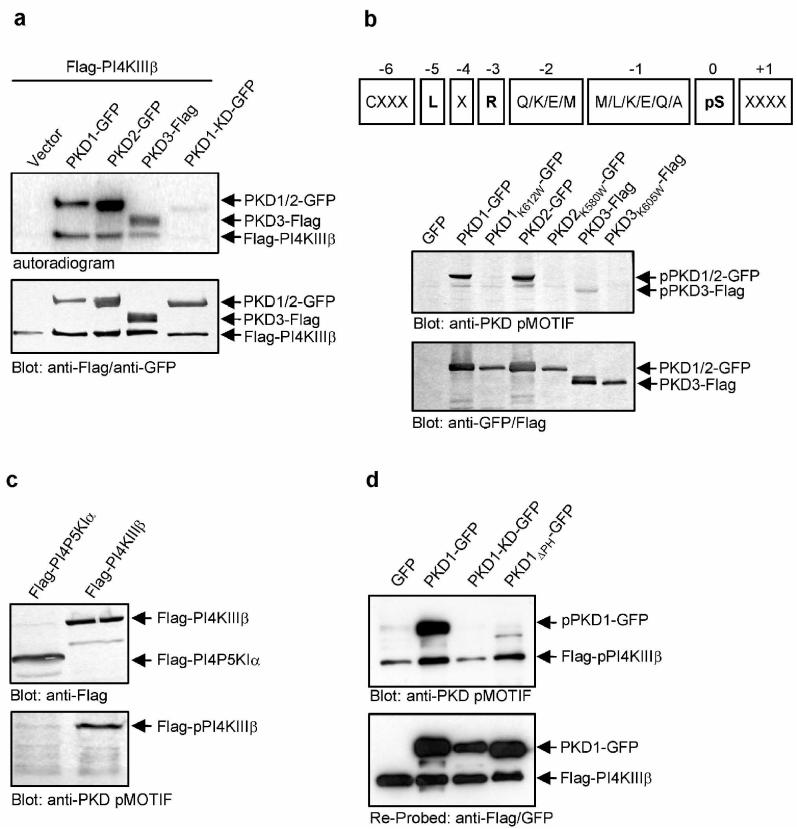
PKD phosphorylates Flag-PI4KIIIβ in vitro and in whole cells.(a) PKD-GFP phosphorylates Flag-PI4KIIIβ in vitro. HEK293 cells were transfected with plasmids encoding the indicated proteins, lysed and PKD-GFP and Flag-PI4KIIIβ were precipitated using anti-GFP and anti-Flag antibodies. Samples were further processed as described in Methods. (b) A PKD pMOTIF antibody is specific for PKD consensus phosphorylation sites. Upper panel: The sequence of the peptide used for immunization of rabbits, derived from the PKD consensus motif is shown. X represents any amino acid, pS represents phospho-serine. Lower panel: HEK293 transfected with the indicated plasmids were lysed and samples were subjected to SDS-PAGE. Proteins were detected using anti-Flag/anti-GFP or anti-PKD pMOTIF antibodies. (c) The PKD pMOTIF antibody specifically detects PI4KIIIβ. HEK293 transfected with the indicated plasmids were lysed and samples were subjected to SDS-PAGE. Proteins were detected using anti-Flag or anti-PKD pMOTIF antibodies. (d) Expression of PKD1-KD-GFP abrogates detection of Flag-PI4KIIIβ with the anti-PKD pMOTIF antibody. HEK293 cells were transfected with the indicated plasmids. Expression of PKD1-GFP and Flag-PI4KIIIβ proteins was controlled by Western Blot using anti-GFP, anti-Flag and anti-PKD pMOTIF antibodies. Results shown are representative of at least three independent experiments.
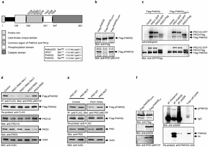
PKD phosphorylates serine 294 in PI4KIIIβ. (a) Schematic view of the domain structure of PI4KIIIβ and the eight known phosphorylation sites and alignment of the putative PKD phosphorylation sites in PI4KIIIβ, serine 294 and 496 with the PKD substrate Kidins220 (bold: critical amino acids: pS: phospho-serine; R: arginine at −3; L/I: critical leucine or isoleucine at −5) (b) the anti-PKD pMOTIF antibody detects the phosphorylated serine 294. HEK293 cells expressing the indicated proteins were lysed and Flag-PI4KIIIβ was detected by Western Blot using anti-Flag and anti-PKD pMOTIF antibodies. (c) PKD phosphorylates serine 294 in Flag-PI4KIIIβ in vitro. HEK293 cells were transfected with plasmids encoding the indicated proteins, lysed and PKD-GFP and Flag-PI4KIIIβ were precipitated using anti-GFP and anti-Flag antibodies. Samples were further processed as described in Methods. (d) Knock-down of PKD1 or PKD2 isoenzymes inhibits Flag-PI4KIIIβ phosphorylation. HEK293E cells were transfected with pSUPER-PKD1-RNAi (2 different sequences; Seq.1 or Seq.2), pSUPER-PKD2-RNAi or pSUPER-PKD3-RNAi. Flag-PI4KIIIβ was precipitated with anti-Flag M2 antibodies and analyzed for phosphorylation at Ser294 (anti-PKD pMOTIF). The blot was re-probed against Flag-PI4KIIIβ. Silencing of endogenous PKD1/2 or PKD3 was monitored by immunoblot analysis with anti-PKD antibodies. Detection of Actin served as a loading control. (e) A PKD mutant with silent mutations in the RNAi-targeted sequence rescues PKD-mediated PI4KIIIβ phosphorylation. HEK293E cells were transfected with pSUPER (control) or pSUPER-PKD1-RNAi. After 24hr cells were transfected with Flag-PI4KIIIβ, combined with PKD1.WT (wild-type) or a PKD1 mutant with two silent mutations in the PKD1-RNAi-targeted sequence (PKD1.MUT). Flag-PI4KIIIβ was precipitated with anti-Flag M2 antibodies and analyzed for phosphorylation at Ser294 (anti-PKD pMOTIF). The blot was re-probed against Flag-PI4KIIIβ. Silencing of endogenous PKD1 was monitored by immunoblot analysis with anti-PKD antibodies. Detection of Actin served as a loading control. (f) A phospho-antibody specific for pS294 in PI4KIIIβ detects endogenous PI4KIIIβ. Endogenous PI4KIIIβ was precipitated from HEK293 cells either with the polyclonal PI4KIIIβ antibody or the polyclonal pS294 serum and detected with the polyclonal pS294 serum. The blot was reprobed with a monoclonal PI4KIIIβ antibody. The preserum served as a control.
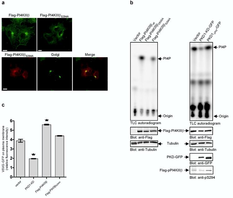
Phosphorylation of PI4KIIIβ at serine 294 is essential for lipid kinase activity and important for transport of VSVG-GFP. (a) Mutation at serine 294 does not affect Golgi localisation. Upper panel: COS7 cells expressing either Flag-PI4KIIIβ or Flag-PI4KIIIβS294A were labelled with antibodies against PI4KIIIβ (green). Lower panel: COS7 cells expressing Flag-PI4KIIIβS294A variants and an YFP-Golgi marker (green) were labelled with antibodies against PI4KIIIβ (red) and analyzed by confocal microscopy. Shown are parallel projections of stacks taken with 0.5 μm width in z-direction. Scale bar represents 10 μm. (b) Phosphorylation at serine 294 affects lipid kinase activity. HEK293 cells were transfected with Flag-PI4KIIIβ variants (left panel) or PKD1-GFP variants and Flag-PI4KIIIβ wild-type (right panel). Flag-PI4KIIIβ variants were precipitated with Flag-antibodies and subjected to lipid kinase assay using phosphatidylinositol (PI) as a substrate. Shown is one representative experiment (n=4). To control the expression of the proteins Western Blots of total cell lysates were performed using anti-Flag, anti-GFP and anti-Tubulin antibodies. The amount of radioactive labelled PI(4)P was normalized to Flag-PI4KIIIβ protein levels. (c) Expression of Flag-PI4KIIIβS294A fails to enhance secretory transport of VSVGGFP protein. HEK293 cells were cotransfected with VSVG-GFP and the indicated plasmids. The amount of VSVG-GFP protein on the plasma membrane is shown as the ratio of red fluorescence (phycoerythrin, staining of plasma membrane localized VSVG protein by indirect immunofluorescence with the 8G5F11 antibody) to green fluorescence (GFP, total VSVG protein expressed). Results are representative (± SEM) of two independent experiments performed. Significance of changes were analysed by student's t-test and indicated by an asterisk (*p < 0.05).
Comment in
-
Signalling for secretion.Nat Cell Biol. 2005 Sep;7(9):851-3. doi: 10.1038/ncb0905-851. Nat Cell Biol. 2005. PMID: 16136181 No abstract available.
References
-
- Liljedahl M, et al. Protein kinase D regulates the fission of cell surface destined transport carriers from the trans-Golgi network. Cell. 2001;104:409–420. - PubMed
-
- Godi A, et al. ARF mediates recruitment of PtdIns-4-OH kinase-beta and stimulatessynthesis of PtdIns(4,5)P2 on the Golgi complex. Nat. Cell Biol. 1999;1:280–287. - PubMed
-
- Rykx A, et al. Protein kinase D: a family affair. FEBS Lett. 2003;546:81–86. - PubMed
Publication types
MeSH terms
Substances
Grants and funding
LinkOut - more resources
Full Text Sources
Other Literature Sources
Molecular Biology Databases
Research Materials
Miscellaneous
