

Organ-specific roles for transcription factor NF-kappaB in reovirus-induced apoptosis and disease
- PMID: 16100570
- PMCID: PMC1184036
- DOI: 10.1172/JCI22428
Organ-specific roles for transcription factor NF-kappaB in reovirus-induced apoptosis and disease
Abstract
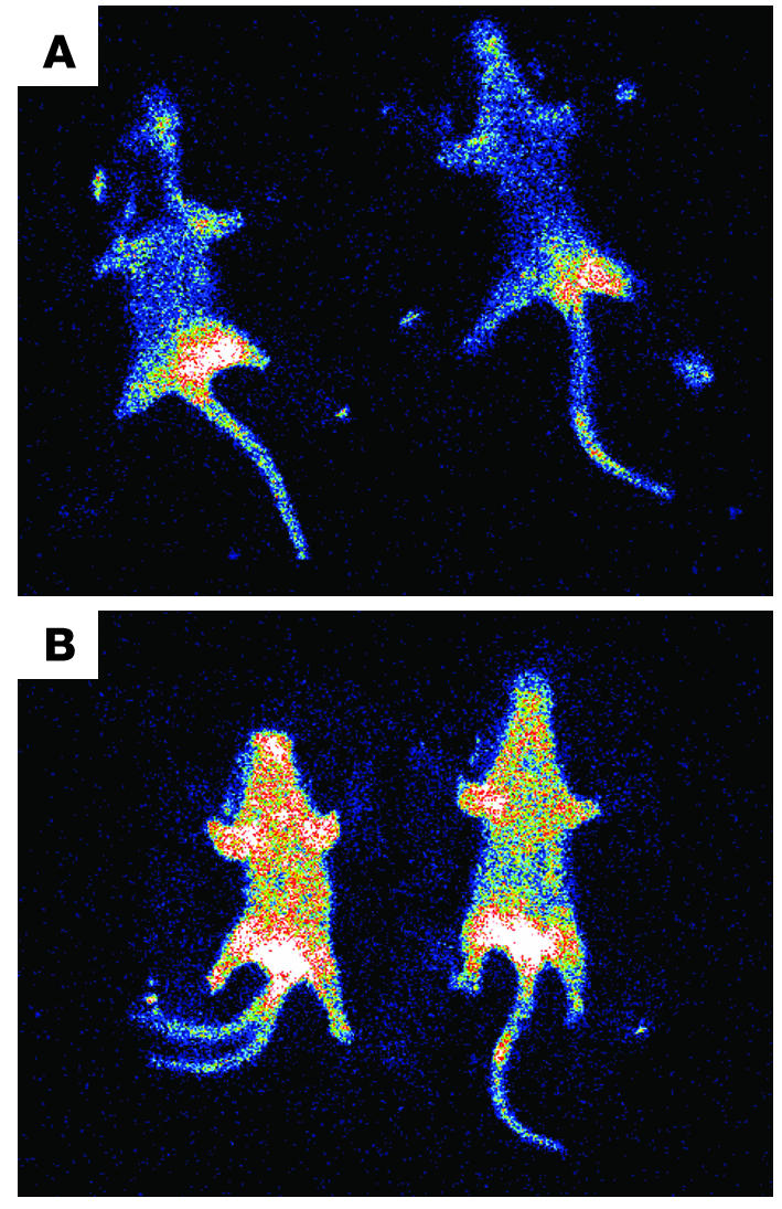
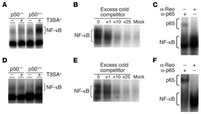
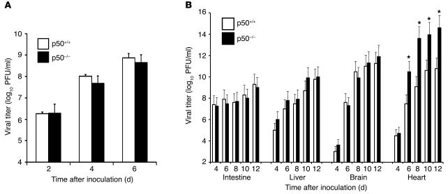
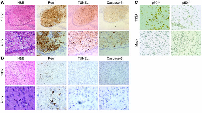
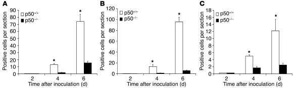
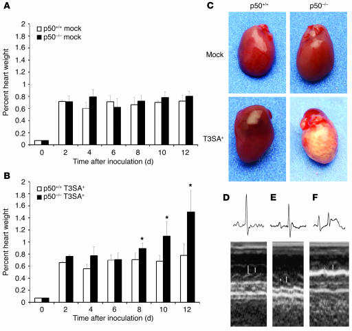
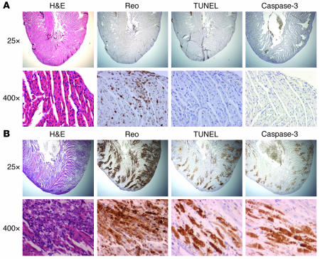
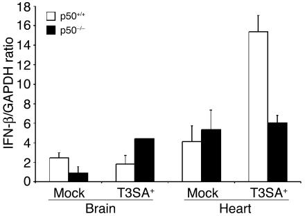
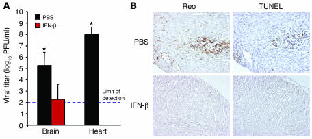
Reovirus induces apoptosis in cultured cells and in vivo. In cell culture models, apoptosis is contingent upon a mechanism involving reovirus-induced activation of transcription factor NF-kappaB complexes containing p50 and p65/RelA subunits. To explore the in vivo role of NF-kappaB in this process, we tested the capacity of reovirus to induce apoptosis in mice lacking a functional nfkb1/p50 gene. The genetic defect had no apparent effect on reovirus replication in the intestine or dissemination to secondary sites of infection. In comparison to what was observed in wild-type controls, apoptosis was significantly diminished in the CNS of p50-null mice following reovirus infection. In sharp contrast, the loss of p50 was associated with massive reovirus-induced apoptosis and uncontrolled reovirus replication in the heart. Levels of IFN-beta mRNA were markedly increased in the hearts of wild-type animals but not p50-null animals infected with reovirus. Treatment of p50-null mice with IFN-beta substantially diminished reovirus replication and apoptosis, which suggests that IFN-beta induction by NF-kappaB protects against reovirus-induced myocarditis. These findings reveal an organ-specific role for NF-kappaB in the regulation of reovirus-induced apoptosis, which modulates encephalitis and myocarditis associated with reovirus infection.
Figures
Similar articles
-
Reovirus-induced apoptosis requires activation of transcription factor NF-kappaB.J Virol. 2000 Apr;74(7):2981-9. doi: 10.1128/jvi.74.7.2981-2989.2000. J Virol. 2000. PMID: 10708412 Free PMC article.
-
Reovirus induction of and sensitivity to beta interferon in cardiac myocyte cultures correlate with induction of myocarditis and are determined by viral core proteins.J Virol. 1998 Feb;72(2):1314-23. doi: 10.1128/JVI.72.2.1314-1323.1998. J Virol. 1998. PMID: 9445032 Free PMC article.
-
Apoptosis induced by mammalian reovirus is beta interferon (IFN) independent and enhanced by IFN regulatory factor 3- and NF-κB-dependent expression of Noxa.J Virol. 2012 Feb;86(3):1650-60. doi: 10.1128/JVI.05924-11. Epub 2011 Nov 16. J Virol. 2012. PMID: 22090144 Free PMC article.
-
Reovirus receptors, cell entry, and proapoptotic signaling.Adv Exp Med Biol. 2013;790:42-71. doi: 10.1007/978-1-4614-7651-1_3. Adv Exp Med Biol. 2013. PMID: 23884585 Free PMC article. Review.
-
Mechanisms of reovirus-induced cell death and tissue injury: role of apoptosis and virus-induced perturbation of host-cell signaling and transcription factor activation.Viral Immunol. 2005;18(1):89-115. doi: 10.1089/vim.2005.18.89. Viral Immunol. 2005. PMID: 15802955 Free PMC article. Review.
Cited by
-
TLR3-mediated NF-{kappa}B signaling in human esophageal epithelial cells.Am J Physiol Gastrointest Liver Physiol. 2009 Dec;297(6):G1172-80. doi: 10.1152/ajpgi.00065.2009. Epub 2009 Sep 24. Am J Physiol Gastrointest Liver Physiol. 2009. PMID: 19779021 Free PMC article.
-
Enter the kill zone: initiation of death signaling during virus entry.Virology. 2011 Mar 15;411(2):316-24. doi: 10.1016/j.virol.2010.12.043. Epub 2011 Jan 22. Virology. 2011. PMID: 21262519 Free PMC article. Review.
-
Nuclear factor-kappaB regulates estrogen receptor-alpha transcription in the human heart.J Biol Chem. 2009 Sep 11;284(37):24705-14. doi: 10.1074/jbc.M109.000463. Epub 2009 Jul 6. J Biol Chem. 2009. PMID: 19584059 Free PMC article.
-
NF-κB activation is cell type-specific in the heart.Virology. 2017 Feb;502:133-143. doi: 10.1016/j.virol.2016.12.022. Epub 2016 Dec 30. Virology. 2017. PMID: 28043025 Free PMC article.
-
Cell Killing by Reovirus: Mechanisms and Consequences.Curr Top Microbiol Immunol. 2023;442:133-153. doi: 10.1007/82_2020_225. Curr Top Microbiol Immunol. 2023. PMID: 32986138 Free PMC article. Review.
References
-
- Tyler, K.L., and Nathanson, N. 2001. Pathogenesis of viral infections. In Fields virology. D.M. Knipe and P.M. Howley, editors. Lippincott-Raven. Philadelphia, Pennsylvania, USA. 199–243.
-
- Tyler, K.L. 2001. Mammalian reoviruses. In Fields virology. D.M. Knipe and P.M. Howley, editors. Lippincott-Raven. Philadelphia, Pennsylvania, USA. 1729–1945.
-
- Virgin, H.W., Tyler, K.L., and Dermody, T.S. 1997. Reovirus. In Viral pathogenesis. N. Nathanson, editor. Lippincott-Raven. New York, New York, USA. 669–699.
-
- Weiner HL, Powers ML, Fields BN. Absolute linkage of virulence and central nervous system tropism of reoviruses to viral hemagglutinin. J. Infect. Dis. 1980;141:609–616. - PubMed
Publication types
MeSH terms
Substances
Grants and funding
LinkOut - more resources
Full Text Sources
Other Literature Sources
Molecular Biology Databases
Research Materials
Miscellaneous
