. 2005 Sep;115(9):2382-92.
doi: 10.1172/JCI23126. Epub 2005 Aug 11.

Involvement of Foxo transcription factors in angiogenesis and postnatal neovascularization

Affiliations

Involvement of Foxo transcription factors in angiogenesis and postnatal neovascularization

Michael Potente et al. J Clin Invest. 2005 Sep.

Abstract

Forkhead box O (Foxo) transcription factors are emerging as critical transcriptional integrators among pathways regulating differentiation, proliferation, and survival, yet the role of the distinct Foxo family members in angiogenic activity of endothelial cells and postnatal vessel formation has not been studied. Here, we show that Foxo1 and Foxo3a are the most abundant Foxo isoforms in mature endothelial cells and that overexpression of constitutively active Foxo1 or Foxo3a, but not Foxo4, significantly inhibits endothelial cell migration and tube formation in vitro. Silencing of either Foxo1 or Foxo3a gene expression led to a profound increase in the migratory and sprout-forming capacity of endothelial cells. Gene expression profiling showed that Foxo1 and Foxo3a specifically regulate a nonredundant but overlapping set of angiogenesis- and vascular remodeling-related genes. Whereas angiopoietin 2 (Ang2) was exclusively regulated by Foxo1, eNOS, which is essential for postnatal neovascularization, was regulated by Foxo1 and Foxo3a. Consistent with these findings, constitutively active Foxo1 and Foxo3a repressed eNOS protein expression and bound to the eNOS promoter. In vivo, Foxo3a deficiency increased eNOS expression and enhanced postnatal vessel formation and maturation. Thus, our data suggest an important role for Foxo transcription factors in the regulation of vessel formation in the adult.


Figures

Figure 1
Figure 1
Foxo1 and Foxo3a are the predominant Foxo transcription factors in endothelial cells. (A) Statistical summary of the Foxo expression profile as assessed in a microarray analysis of total RNA isolated from HUVECs. Data are presented as mean ± SEM; n = 4. The values on the y axis represent a ratio normalized to the mean fluorescent intensity of all genes on the chip. (B) Cell fractions were prepared from HUVECs that were either left untreated, serum starved for 24 hours, or transfected with Flag-Foxo1, HA-Foxo3a, or HA-Foxo4 and subjected to SDS-PAGE. Western blot analysis was performed using the indicated antibodies. Tagged, epitope-tagged; endo., endogenous. (C) HUVECs were serum-starved for 24 hours and then either left untreated or stimulated with VEGF (50 ng/ml) for the indicated times. Cell lysates were subjected to Western blot analysis with antibodies against phospho-Foxo1 (Thr 24), phospho-Foxo3a (Thr 32), or total Foxo1. (D) HUVECs were serum starved for 24 hours, then incubated with 10 μM LY294002 for 1 hour and stimulated with VEGF (50 ng/ml) for the indicated times. Cell lysates were analyzed by Western blot analysis using an antibody directed against phospho-Foxo1. The total level of protein was assayed by Western blot analysis using an anti-Foxo1 and anti-Akt antibody. FCS (20%) was used as a positive control.
Figure 2
Figure 2
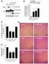
Overexpression of a gain-of-function mutant of Foxo1 or Foxo3a inhibits endothelial sprout formation and migration. (A) HUVECs were transfected with constitutively active Foxo1, Foxo3a, Foxo4, or mock control. Twenty-four hours later, cells were lysed and subjected to Western blot analysis with antibodies against Flag and HA. An antibody directed against tubulin was used as loading control. (B) HUVECs were transfected with a forkhead-responsive element reporter construct (6xDBE) along with plasmids encoding either constitutively active Foxo1, Foxo3a, or Foxo4. A transfected empty vector (pcDNA) was used a control. At 24 hours after transfection, cells were lysed, and luciferase relative to renilla luciferase activity was measured. × Control, fold value relative to pcDNA-transfected cells. The statistical summary represents the mean ± SEM; n = 3. (C and D) Statistical summary and representative micrographs of the tube-forming activity. HUVECs were seeded on Matrigel Basement Membrane Matrix 18 hours after transfection with the indicated plasmids. The length of capillary-like structures was measured by light microscopy after 24 hours in a blinded fashion. Data are presented as mean ± SEM; n = 5 (Foxo1), n = 6 (Foxo3a), n = 5 (Foxo4). *P < 0.001 versus control. Magnification, ×50. (E) HUVECs were transfected with the constitutively active constructs of Foxo1, Foxo3a, and Foxo4 and were seeded in the upper chamber of a modified Boyden chamber 18 hours after transfection. Endothelial cell migration was assessed using VEGF (50 ng/ml) as chemoattractant after 24 hours of incubation. Data are presented as mean ± SEM. **P < 0.05 versus control; n = 3.
Figure 3
Figure 3
Silencing Foxo1 or Foxo3a activity enhances the angiogenic activity of endothelial cells. (A) HUVECs were transfected with 2 different siRNAs targeted against either Foxo1 (Foxo1 I and Foxo1 II) or Foxo3a (Foxo3a I and Foxo3a II). A nonrelated scrambled siRNA was used a control. Cell lysates were subjected to Western blotting using antibodies against Foxo1 and Foxo3a. Tubulin was used a loading control. (B and C) HUVECs were transfected with 2 different siRNAs targeted against Foxo1, Foxo3a, or a scrambled oligonucleotide. After 18 hours, cells were seeded on a Growth Factor Reduced Matrigel in the presence of VEGF (50 ng/ml). Cumulative sprout length of capillary-like structures was measured by light microscopy after 24 hours. Representative micrographs and statistical summary are shown. Data are presented as mean ± SEM; n = 4 (Foxo1), n = 6 (Foxo3a). *P < 0.001 versus control. (D) Representative micrographs and statistical summary of endothelial cells transfected with siRNAs targeted against Foxo1, Foxo3a, or scrambled control. After 18 hours, cells were seeded in the upper chamber of modified Boyden chamber. Endothelial cell migration was assessed using VEGF (50 ng/ml) as chemoattractant. After 24 hours, nonmigrating cells on the upper side of the chamber were mechanically removed, and the remaining cells on the lower side were fixed and stained with DAPI. Data are presented as mean ± SEM; n = 5. *P < 0.001 versus control. (E) Three-dimensional in vitro angiogenesis with collagen gel–embedded spheroids of Foxo1, Foxo3a, or scrambled siRNA–transfected endothelial cells. Cumulative length of all sprouts originating from an individual spheroid was quantified after 24 hours. Statistical summary represents the mean ± SEM; n = 3. **P < 0.05 versus control. Magnification: ×50 (B); ×200 (D); ×100 (E).
Figure 4
Figure 4
Transcriptome of endogenous Foxo1 and Foxo3a in endothelial cells. HUVECs were transfected with a Foxo1- and Foxo3a-specific siRNA or a scrambled oligonucleotide siRNA (each n = 3). Total RNA was isolated after 24 hours, and the gene expression profile was assessed with the Affymetrix gene chip expression assay. (A) Gene tree analysis of selected angiogenesis-related genes. Red indicates high expression. Genes regulated by Foxo1 and Foxo3a siRNA transfection are indicated by white boxes. (B) Validation of angiogenesis-related Foxo1- and Foxo3a-regulated genes. HUVECs were transfected with Foxo1- and Foxo3a-specific siRNAs and lysed 24 hours after transfection. Cell lysates were subjected to SDS-PAGE, and expression of eNOS, Elk-3, Ang2, Foxo1, and Foxo3a was determined by Western blot analysis. Tubulin and ERK1/2 served as loading controls.
Figure 5
Figure 5
Foxo1 and Foxo3a are transcriptional repressors of eNOS. (A) eNOS expression in HUVECs that were transfected with constitutively active constructs of Flag–Foxo1 A3 and HA–Foxo3a A3. Cell lysates were prepared at the indicated time points, and expression of eNOS, Flag, HA, and tubulin was determined by Western blotting using the respective antibodies. (B) eNOS expression in HUVECs that were transfected with Foxo1- or Foxo3a-specific siRNA and lysed at the indicated time points. eNOS, Foxo1, and Foxo3a expression was determined by immunoblotting. SCR, scrambled siRNA; FX1, Foxo1 siRNA; FX3a, Foxo3a siRNA. (C) eNOS expression in the aorta of Foxo3a+/+ and Foxo3a–/– mice. After the aortas from the indicated groups of mice were removed, cell lysates were prepared, and expression of eNOS was determined by immunoblotting. Equal protein loading was confirmed with a tubulin antibody. (D) HUVECs were transfected with Foxo1 A3 (Flag-Foxo1 A3), Foxo3a A3 (HA–Foxo3a A3), Foxo4 A3 (HA–Foxo4 A3), or vector control. After 24 hours, chromatin-bound DNA was immunoprecipitated with an antibody against the Flag or HA epitope. Immunoprecipitated DNA was analyzed by PCR using a primer combination that encompassed the forkhead responsive element (FHRE). The pGL3-eNOS-3500 plasmid was used as a positive control for the PCR. eNOS primers for the eNOS coding sequence were used as a negative control to exclude nonspecific precipitated DNA. CDS, coding sequence. (E) HUVECs were cotransfected with pGL3-eNOS-3500 and either Foxo1 A3 or the empty vector pcDNA3 (pcDNA). Luciferase activity was measured 24 hours later. Values are expressed as the level of luciferase activity of Foxo1 A3 relative to that of pcDNA, which was set as 100%. Data are presented as mean ± SEM; n = 6. *P < 0.001.
Figure 6
Figure 6
Foxo3a modulates neovascularization capacity in vivo. (A) Foxo3a+/+ and Foxo3a–/– mice were subjected to hind limb ischemia, and perfusion was assessed 14 days after onset of ischemia using laser Doppler imaging. Low or no perfusion is shown as dark blue, whereas the highest perfusion level is shown as red. Arrows indicate the ischemic leg. Quantitative results are presented as mean ± SEM; n = 8. *P = 0.002. (B) Capillary density (ratio of the number of capillaries to the number of myocytes) was determined in 8-μm frozen sections of the adductor and semimembraneous muscles. Quantitative results are presented as mean ± SEM; n = 8 (Foxo3a+/+), n = 7 (Foxo3a–/–). (C) Conductance vessels in the adductor and semimembraneous muscles were identified by size (>20 μm) and smooth muscle actin staining using a Cy3-labeled mouse monoclonal antibody for smooth muscle actin. The number of small (<50 μm), medium (50–100 μm), and large vessels was determined separately. Data are presented as mean ± SEM; n = 6 (Foxo3a+/+), n = 5 (Foxo3a–/–). **P = 0.01. (D) Statistical summary and representative micrographs of blood vessel infiltration in Matrigel sections stained with a smooth muscle actin antibody in wild-type and Foxo3a–/– mice. Quantitative results are presented as mean ± SEM; n = 7 (Foxo3a+/+), n = 8 (Foxo3a–/–). Scale bars in C and D, 100 μm.

Similar articles

Cited by

References

    1. Tran H, Brunet A, Griffith EC, Greenberg ME. The many forks in FOXO’s road [review] Sci STKE. 2003;2003:RE5. - PubMed
    1. Burgering BM, Medema RH. Decisions on life and death: FOXO Forkhead transcription factors are in command when PKB/Akt is off duty [review] J. Leukoc. Biol. 2003;73:689–701. - PubMed
    1. Accili D, Arden KC. FoxOs at the crossroads of cellular metabolism, differentiation, and transformation. Cell. 2004;117:421–426. - PubMed
    1. Kitamura T, et al. The forkhead transcription factor Foxo1 links insulin signaling to Pdx1 regulation of pancreatic β cell growth. J. Clin. Invest. 2002;110:1839–1847. doi:10.1172/JCI200216857. - PMC - PubMed
    1. Castrillon DH, Miao L, Kollipara R, Horner JW, DePinho RA. Suppression of ovarian follicle activation in mice by the transcription factor Foxo3a. Science. 2003;301:215–218. - PubMed

Publication types

MeSH terms