. 2006 Jan 1;393(Pt 1):141-50.
doi: 10.1042/BJ20050327.

SMAD 8 binding to mice Msx1 basal promoter is required for transcriptional activation

Affiliations

SMAD 8 binding to mice Msx1 basal promoter is required for transcriptional activation

Renata Binato et al. Biochem J. .

Abstract

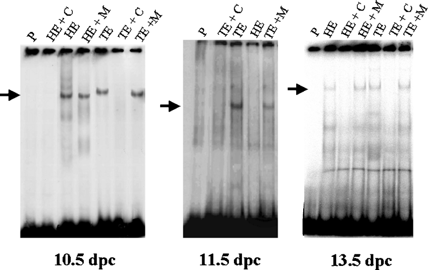
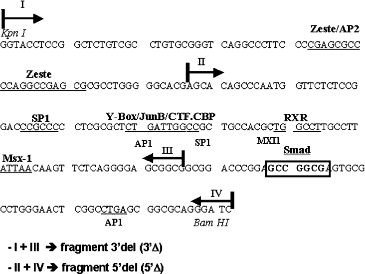
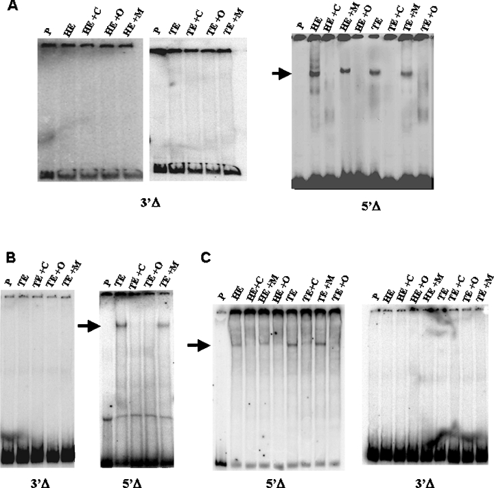
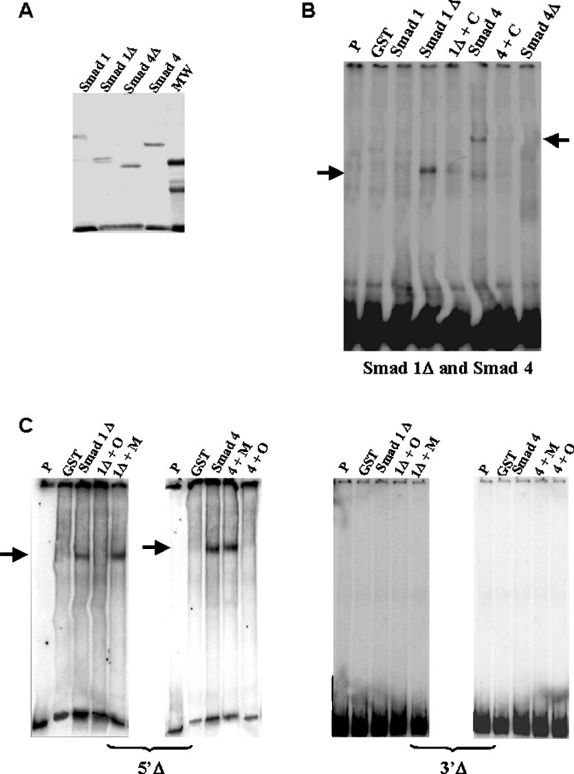
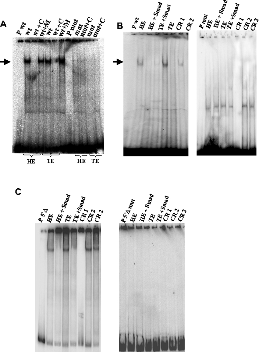
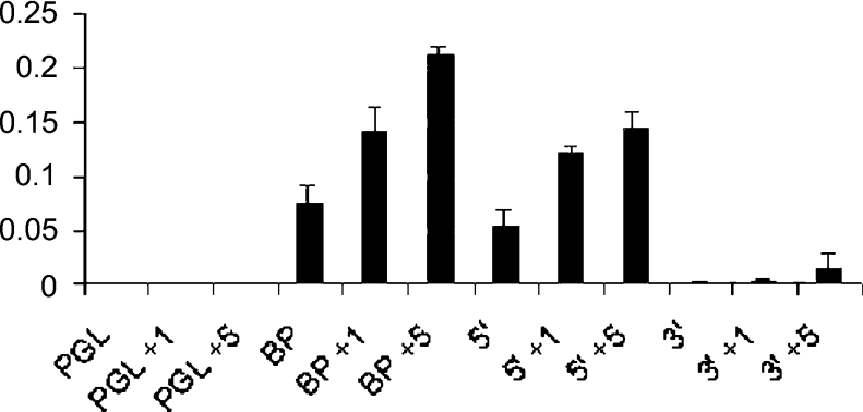
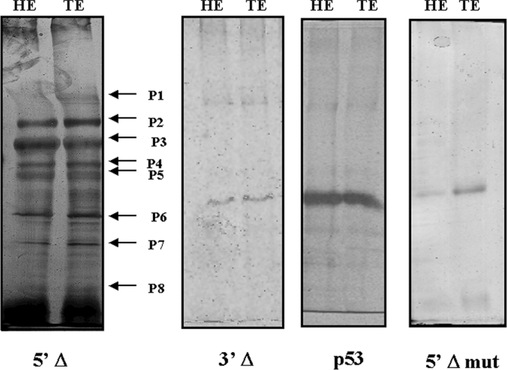
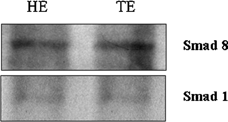
The Msx1 gene in mice has been proven to be induced by BMP (bone morphogenetic protein) proteins, and three binding sites for SMAD, an intracellular BMP signalling transducer, have already been identified in its promoter. Gel shift analyses were performed and they demonstrated that the consensus found very near the transcription start site, a region designed BP (basal promoter), is functional for binding nuclear proteins from 10.5, 11.5 and 13.5 dpc (days post-coitum) embryos. Notably, this binding occurs only when the SMAD-binding consensus sequence is maintained, suggesting that it is required for the formation of a protein complex over BP. Binding of purified SMAD 1 and SMAD 4 as well as supershift assay with SMAD 1/SMAD 5/SMAD 8 antibody proved that a SMAD protein is present in this complex. Transfection assays in cell cultures with fragments from BP driving the expression of luciferase confirmed that only in the presence of the SMAD consensus site is Msx1 expression activated. A proteomic analysis of the complex components after immunoprecipitation identified several proteins necessary to activate transcription including SMAD 8. Our results suggest that BMP2/BMP4 signalling through SMAD 8 is required for transcriptional activation of the mouse Msx1 gene.


Figures

Figure 1
Figure 1. Gel shift analysis of binding reactions using fragment BP as a probe and head (HE) or trunk (TE) embryo extracts from different ages (10.5, 11.5 and 13.5 dpc)
Specificity of DNA–protein complexes is demonstrated by competition reactions using 100 times unlabelled BP (lanes HE+C and TE+C) and by competition reactions using 100 times unlabelled mutant oligonucleotide DNA for the SMAD consensus site (lanes HE+M and TE+M). Each lane indicated as ‘P’ corresponds to migration of the probe alone. The arrows show the specific binding.
Figure 2
Figure 2. Deleted fragments generation scheme
Subfragments from BP were generated by PCR and cloning. One fragment was deleted in 3′-region (3′Δ) and the other was deleted in 5′-region (5′Δ). Note that the SMAD consensus binding site (boxed sequence) is deleted in the 3′Δ construct.
Figure 3
Figure 3. EMSA of head (HE) or trunk (TE) nuclear extracts from embryos at different stages of development, using the deleted fragments described in Figure 2
(A) Gel shift using 10.5 dpc head and trunk nuclear embryo extracts. (B) Gel shift using 11.5 dpc trunk nuclear embryo extract. (C) Gel shift using 13.5 dpc head and trunk nuclear embryo extracts. (AC) The fragment used as a probe is indicated below each gel. Lanes indicated as ‘P’ represent migration of the probes alone. Competition reactions with 100 times unlabelled probe are indicated as ‘+C’. Competition reactions with 100 times unlabelled wild-type oligonucleotide for the SMAD consensus site are indicated by ‘+O’. Competition reactions with 100 times unlabelled mutated oligonucleotide for the SMAD consensus site are indicated as ‘+M’. The arrow shows the specific binding that occurs only with the 5′Δ fragment.
Figure 4
Figure 4. Purified SMAD 1 and SMAD 4 bind to BP and 5′Δ and 3′Δ fragments
(A) Gel of purified recombinant proteins SMAD 1, SMAD 1Δ, SMAD 4 and SMAD 4Δ. MW, molecular mass standards. (B) Gel shift analysis of complexes between GST–SMAD fusion proteins and BP fragment. The protein used is indicated above each lane. Lanes indicated as ‘P’ and ‘GST’ show migration of the probe alone and reaction using GST peptide respectively. We observed binding reactions involving SMAD 1ΔMH2 (called SMAD 1Δ) and SMAD 4 (arrows). Competition reactions are indicated by the symbol ‘+C’ and correspond to addition of 100 times unlabelled BP. (C) Gel shift analysis of purified GST–SMAD 1ΔMH2 and SMAD 4 fusion protein complexes with the 5′Δ and 3′Δ fragments. The fragment used as a probe is indicated below each gel. Lanes indicated as ‘P’ and ‘GST’ show migration of the probe alone and reaction using GST peptide respectively. Competition reactions with 100 times unlabelled wild-type oligonucleotide for the SMAD consensus site are indicated as ‘+O’ and competition reactions with 100 times unlabelled mutated oligonucleotide for the SMAD consensus site are indicated as ‘+M’. The arrows show the specific binding.
Figure 5
Figure 5. SMAD proteins are present in 13.5 dpc embryo nuclear extracts and participate in the protein complex formed with a SMAD consensus oligonucleotide
(A) EMSA performed with head (HE) and trunk (TE) nuclear extracts from 13.5 dpc embryos and the wild-type (wt) or mutant (mut) oligonucleotides for the SMAD consensus binding site. Lanes indicated as ‘P wt’ and ‘P mut’ show migration of the probe alone. Competition reactions are indicated by the symbol ‘+C’ and correspond to the addition of 100 times unlabelled wild-type oligonucleotide. Competition reactions with 100 times unlabelled mutated oligonucleotide are indicated as ‘+M’. The arrows show specific binding confirming that mutations in the SMAD consensus site eliminate binding. (B) Supershift analysis of binding between head and trunk 13.5 dpc embryo nuclear extract and wild-type and mutated oligonucleotide. Lanes indicated as ‘P wt’ and ‘P mut’ show migration of the probe alone. Lanes indicated as HE and TE correspond to incubation of the probe with protein extract from head and trunk respectively. The addition of the SMAD 1/SMAD 5/SMAD 8 antibody in binding reactions with protein extract from head and trunk is analysed in lanes indicated as ‘head+SMAD’ and ‘trunk+SMAD’ respectively. Reaction indicated as CR 1 corresponds to a control reaction without nuclear proteins and CR 2 corresponds to a control reaction using an unspecific antibody (anti-P53). (C) Supershift analysis of binding between head and trunk 13.5 dpc embryo nuclear extract and 5′Δ fragment and 5′Δ mutated fragment. Lanes indicated as ‘P 5′Δ’ and ‘P 5′Δmut’ show migration of the probe alone. Lanes indicated as head and trunk correspond to incubation of the probe with protein extract from head and trunk respectively. The addition of the SMAD 1/SMAD 5/SMAD 8 antibody in binding reactions with protein extract from head and trunk is analysed in lanes indicated as ‘HE+SMAD’ and ‘TE+SMAD’ respectively. Reaction indicated as CR 1 correspond to a control reaction without nuclear proteins and CR 2 corresponds to a control reaction using an unspecific antibody (anti-P53).
Figure 6
Figure 6. P19 cells were transiently transfected using the reporter plasmid PGL-basic2 (PGL – containing the luciferase gene) alone or using the constructions BP, 5′Δ and 3′Δ
The cells were treated or not, 24 h after transfection, with 1 nM (PGL+1) or 5 nM (PGL+5) BMP4 for 5 h. Whole cell lysates were prepared and analysed for luciferase activity. The histogram shows induction of luciferase activity when we used both constructions containing the SMAD consensus binding site (BP and 5′Δ) and this activity is increased in the presence of 1 and 5 nM BMP4 (BP+1 nM, BP+5 nM, 5′Δ+1 nM and 5′Δ+5 nM). In contrast, the levels of luciferase using the construction without the SMAD consensus binding site (3′Δ) were minimal even in the presence of BMP4 (3′Δ+1 nM and 3′Δ+5 nM). PGL, PGL+1 nM and PGL+5 nM indicate the reporter plasmid alone, untreated with BMP and treated with 1 and 5 nM BMP4 respectively. These data are representative with P value <0.001.
Figure 7
Figure 7. SDS/polyacrylamide gel of immunoprecipitation products
Lanes named HE and TE correspond to reactions using 13.5 dpc embryo nuclear protein extracts from head and trunk respectively with anti-SMAD 1/SMAD 5/SMAD 8 antibody. The fragment used as a probe [5′-deleted fragment (5′Δ), 5′Δ mutated fragment (5′Δ mut) and 3′Δ fragment (3′Δ)] is indicated below each gel. The immunoprecipitated complex showed apparently the same composition using nuclear protein extracts from head or trunk. The arrows P1–P8 in the gel using 5′Δ fragment indicate the protein bands analysed by MS. The gel using 3′Δ fragment and 5′Δ mutated fragment did not show any immunoprecipitated protein. A control immunoprecipitation using an unspecific antibody (anti-P53) also did not show any protein.
Figure 8
Figure 8. Western-blot analysis using 13.5 dpc embryo nuclear protein extracts from head (HE) and trunk (TE) with anti-SMAD 1 or anti-SMAD 8 antibodies
The extract and the antibody used are indicated in each gel. The bands showed the presence of each protein in the extracts but in different concentrations.

References

    1. Robert B., Sassoon D., Jacq B., Gehring W., Buckingham M. Hox-7, a mouse homeobox gene with a novel pattern of expression during embryogenesis. EMBO J. 1989;8:91–100. - PMC - PubMed
    1. Hill R. E., Jones P. F., Rees A. R., Sime C. M., Justice M. J., Copeland N. G., Jenkins A., Graham E., Davidson D. R. A new family of mouse homeobox-containing genes:molecular structure, chromosomal location, and developmental expression of Hox-7-1. Genes Dev. 1989;3:26–37. - PubMed
    1. Satokata I., Maas R. Msx-1 deficient mice exhibit cleft palate and abnormalities of craniofacial and tooth development. Nat. Genet. 1994;6:348–356. - PubMed
    1. Houzelstein D., Cohen A., Buckingham M. E., Robert B. Insertional mutation of the mouse Msx-1 homeobox gene by an nLacZ reporter gene. Mech. Dev. 1997;65:123–133. - PubMed
    1. Davidson D. R., Hill R. E. Msh-like genes: a family of homeobox genes with wide ranging expression during vertebrate development. Semin. Dev. Biol. 1991;2:405–412.

Publication types

MeSH terms