. 2005 Aug 15;65(16):7301-9.
doi: 10.1158/0008-5472.CAN-05-1064.

Protein kinase C-epsilon regulates the apoptosis and survival of glioma cells

Affiliations

Protein kinase C-epsilon regulates the apoptosis and survival of glioma cells

Hana Okhrimenko et al. Cancer Res. .

Abstract

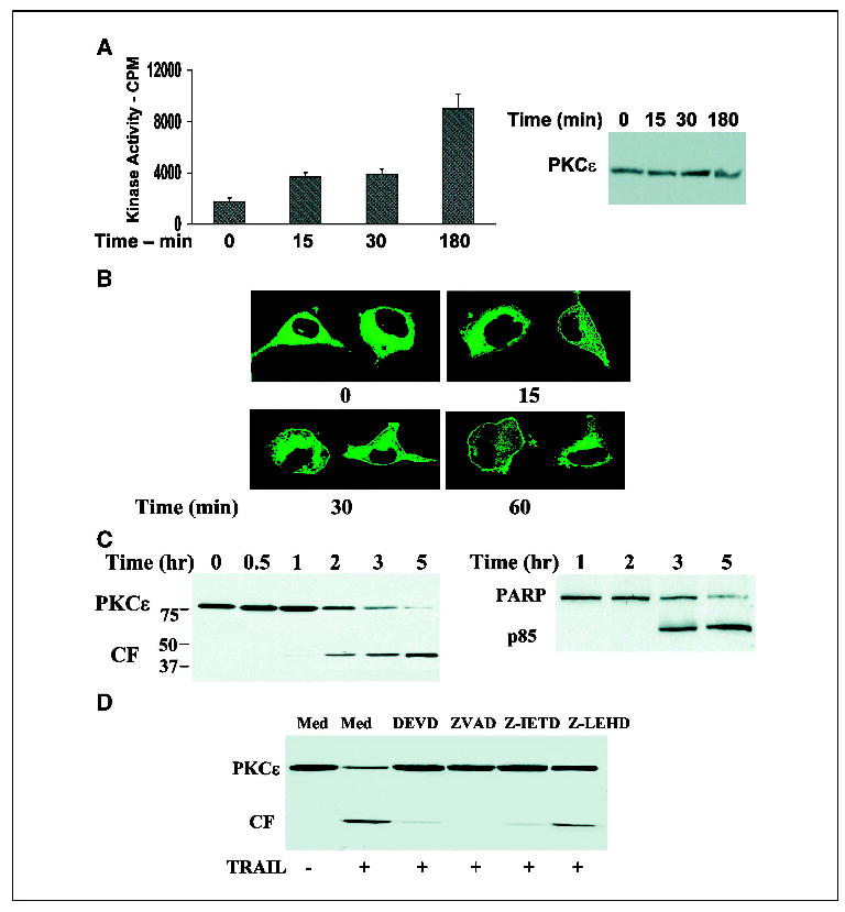
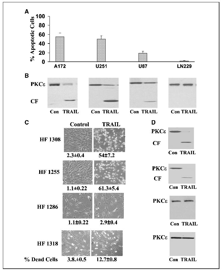
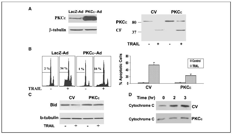
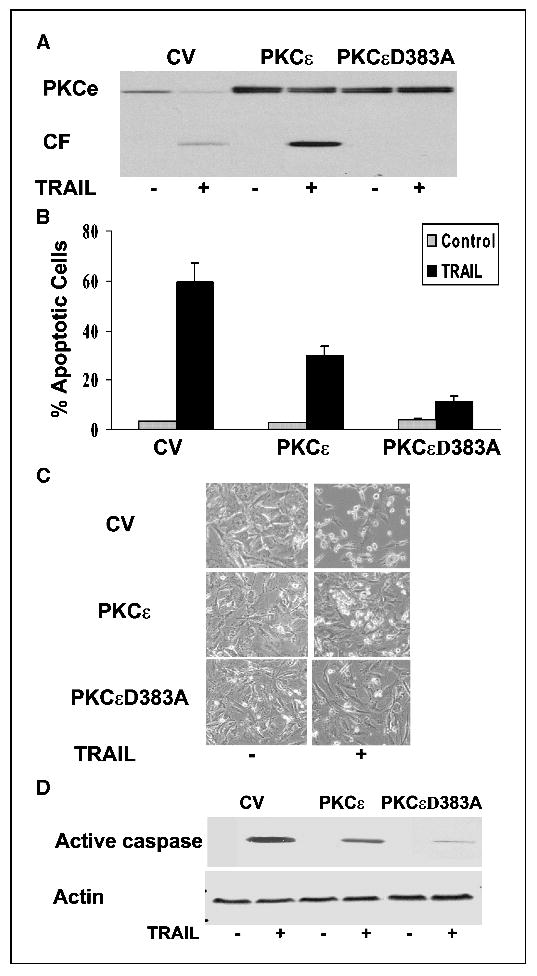
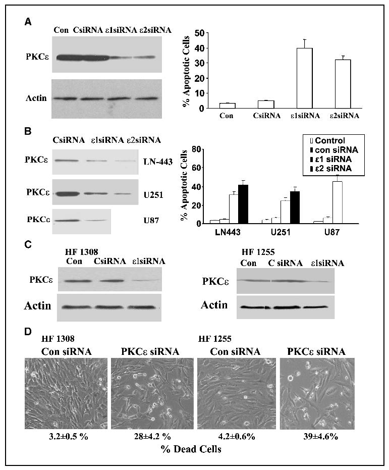
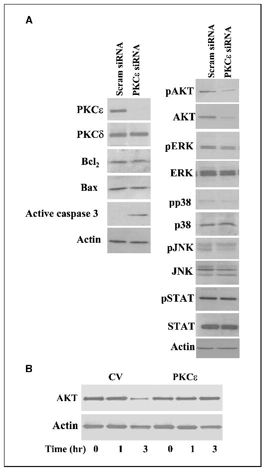
In this study, we examined the role of protein kinase C (PKC)-epsilon in the apoptosis and survival of glioma cells using tumor necrosis factor-related apoptosis inducing ligand (TRAIL)-stimulated cells and silencing of PKCepsilon expression. Treatment of glioma cells with TRAIL induced activation, caspase-dependent cleavage, and down-regulation of PKCepsilon within 3 to 5 hours of treatment. Overexpression of PKCepsilon inhibited the apoptosis induced by TRAIL, acting downstream of caspase 8 and upstream of Bid cleavage and cytochrome c release from the mitochondria. A caspase-resistant PKCepsilon mutant (D383A) was more protective than PKCepsilon, suggesting that both the cleavage of PKCepsilon and its down-regulation contributed to the apoptotic effect of TRAIL. To further study the role of PKCepsilon in glioma cell apoptosis, we employed short interfering RNAs directed against the mRNA of PKCepsilon and found that silencing of PKCepsilon expression induced apoptosis of various glioma cell lines and primary glioma cultures. To delineate the molecular mechanisms involved in the apoptosis induced by silencing of PKCepsilon, we examined the expression and phosphorylation of various apoptosis-related proteins. We found that knockdown of PKCepsilon did not affect the expression of Bcl2 and Bax or the phosphorylation and expression of Erk1/2, c-Jun-NH2-kinase, p38, or STAT, whereas it selectively reduced the expression of AKT. Similarly, TRAIL reduced the expression of AKT in glioma cells and this decrease was abolished in cells overexpressing PKCepsilon. Our results suggest that the cleavage of PKCepsilon and its down-regulation play important roles in the apoptotic effect of TRAIL. Moreover, PKCepsilon regulates AKT expression and is essential for the survival of glioma cells.


Figures

Figure 1
Figure 1
TRAIL induces activation, cleavage, and down-regulation of PKCɛ in glioma cells. A172 cells were treated with TRAIL for 0 to 3 hours and the activity of PKCɛ was determined using an immune complex PKCɛ kinase assay. Samples were also subjected to immunoblot analysis for the determination of total PKCɛ levels (A). Translocation of PKCɛ in response to TRAIL was determined in A172 cells transfected with PKCɛ-GFP. Cells were stimulated with TRAIL (100 ng/mL) for various periods of time and were then visualized by confocal microscopy (B). Cleavage and expression of PKCɛ was determined in A172 cells treated with TRAIL for 0 to 5 hours by Western blot using an anti-PKCɛ antibody that recognizes the catalytic domain [anti-PKCɛ (C-15), Santa Cruz] and cell apoptosis was measured in parallel using PARP cleavage (C). The role of caspase 3, 8, and 9 in the cleavage of PKCɛ was determined using pretreatment of the cells with DEVD, ZVAD, Z-IRTD, or Z-LEHD (10 μmol/L) for 30 minutes followed by incubation with TRAIL for an additional 3 hours (D). Columns, means; bars, ±SE (A); results from one of four separate experiments which gave similar results (B, C, and D).
Figure 2
Figure 2
Cleavage and down-regulation of PKCɛ in TRAIL-sensitive and -resistant glioma cells. Various glioma cell lines (A and B) and primary glioma cultures (C and D) were treated with 100 ng/mL TRAIL for 5 hours. Cell apoptosis was determined using propidium iodide staining and fluorescence-activated cell sorting analysis (A) or by trypan blue exclusion assay (C) and the expression and cleavage of PKCɛ was determined using Western blot analysis (B and D). The morphology of the cells was monitored under a phase contrast light microscope (C). Columns, means; bars, ±SE (A); results from one representative experiment out of four similar experiments (B, C, and D).
Figure 3
Figure 3
Overexpression of PKCɛ protects A172 cells from TRAIL-induced apoptosis. A172 cells were infected with adenovirus vectors expressing PKCɛ (PKCɛ-AdV) and LacZ (CV, LacZ-AdV; A and B) or transfected with control vector or tg2b-PKCɛ (A and B). Following 24 hours, the cells were treated with TRAIL for 5 hours, cleavage of PKCɛ was determined using Western blot analysis (A) and cell apoptosis was determined using propidium iodide staining and fluorescence-activated cell sorting analysis (B). Bid expression was determined after 90 minutes (C) and cytochrome c release was determined after 2 hours of treatment (D) as described in Materials and Methods. The results are representative of four similar experiments (A, B, C, and D); columns, means; bars, ±SE (B).
Figure 4
Figure 4
Cleavage of PKCɛ plays a role in the apoptotic effect of TRAIL. A172 cells were transfected with control vector, PKCɛ, or PKCɛD383A. Following 48 hours, the cells were treated with TRAIL for 5 hours and the cleavage of PKCɛ was determined using Western blot analysis (A). Cell apoptosis was determined using propidium iodide staining and fluorescence-activated cell sorting analysis (B). The cells were also visualized using a phase contrast microscope (C). The levels of active caspase 3 were determined using Western blot analysis (D). The results are representative of four similar experiments (A, C, and D); columns, means; bars, ±SE (B).
Figure 5
Figure 5
Silencing of PKCɛ expression induces apoptosis in glioma cells. The glioma cell lines, A172 (A) LN-443, U251, and U87 (B) and the primary glioma cultures HF1308 and HF1255 (C and D) were transfected with 50 nmol/L scrambled siRNA (CsiRNA) and siRNAs targeting the mRNA of PKCɛ (PKCɛ siRNAs). Following 72 hours, the expression of PKCɛ was determined using Western blot analysis (A, B, and C) and cell apoptosis was determined using propidium iodide staining and fluorescence-activated cell sorting analysis (A and B) or trypan blue exclusion and phase contrast microscopy (D). The results represent one of four separate experiments, which gave similar results (A and B) or are the means ±SE of five independent experiments (A and B; FACS analysis).
Figure 6
Figure 6
Loss of PKCɛ reduces the expression of AKT in glioma cells. A172 cells were transfected with 50 nmol/L scrambled siRNA or with siRNAs targeting the mRNA of PKCɛ (PKCɛ siRNA). Following 72 hours, the expression of PKCɛ, PKCδ, active caspase 3, Bax and Bcl2 as well as the expression and phosphorylation of AKT, Erk1/2, p38, JNK, and STAT1 were determined using Western blot analysis (A). To examine the role of PKCɛ in AKT expression in TRAIL-treated cells, A172 cells overexpressing control vector or PKCɛ were treated with TRAIL (100 mg/mL) for 1 to 3 hours and the expression of AKT was measured using Western blot analysis (B). The results represent one of four separate experiments, which gave similar results.

References

    1. Nishizuka Y. The molecular heterogeneity of protein kinase C and its implications for cellular regulation. Nature. 1988;334:661–5. - PubMed
    1. Hug H, Sarre TF. Protein kinase C isoenzymes: divergence in signal transduction? Biochem J. 1993;291:329–43. - PMC - PubMed
    1. Gutcher I, Webb PR, Anderson NG. The isoform-specific regulation of apoptosis by protein kinase C. Cell Mol Life Sci. 2003;60:1061–70. - PMC - PubMed
    1. Ruvolo PP, Deng X, Carr BK, May WS. A functional role for mitochondrial protein kinase Cα in Bcl2 phosphorylation and suppression of apoptosis. J Biol Chem. 1998;273:25436–42. - PubMed
    1. Jamieson L, Carpenter L, Biden TJ, Fields AP. Protein kinase Cι activity is necessary for Bcr-Abl-mediated resistance to drug-induced apoptosis. J Biol Chem. 1999;274:3927–30. - PubMed

Publication types

MeSH terms