. 2005 Oct 14;280(41):34530-7.
doi: 10.1074/jbc.M508890200. Epub 2005 Aug 15.

Involvement of the p97-Ufd1-Npl4 complex in the regulated endoplasmic reticulum-associated degradation of inositol 1,4,5-trisphosphate receptors

Affiliations

Involvement of the p97-Ufd1-Npl4 complex in the regulated endoplasmic reticulum-associated degradation of inositol 1,4,5-trisphosphate receptors

Kamil J Alzayady et al. J Biol Chem. .

Abstract

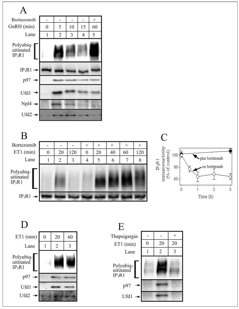
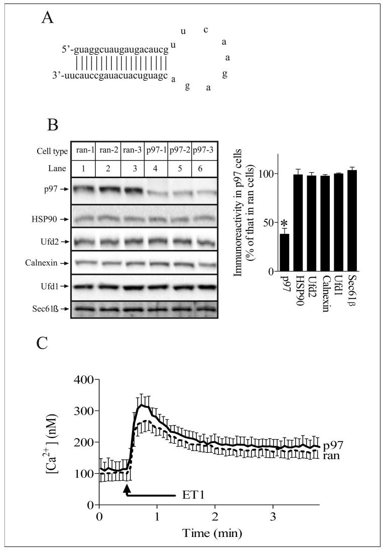
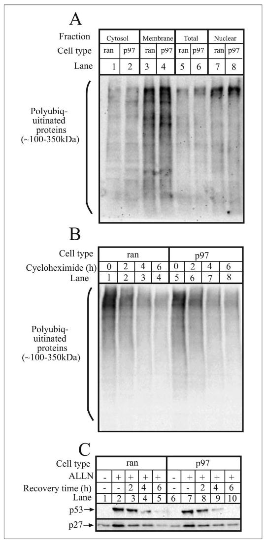
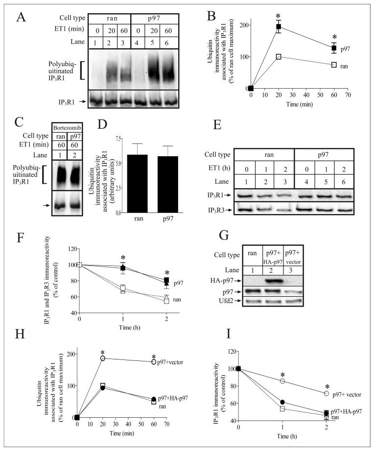
Inositol 1,4,5-trisphosphate (IP(3)) receptors form tetrameric, IP(3)-gated channels in endoplasmic reticulum membranes that govern the release of Ca(2+) from this organelle. In response to activation of certain G protein-coupled receptors that persistently elevate IP(3) concentration, IP(3) receptors are ubiquitinated and degraded by the ubiquitin-proteasome pathway. IP(3) receptor ubiquitination is mediated by the ubiquitin-conjugating enzyme, (mam)Ubc7, a component of the endoplasmic reticulum-associated degradation pathway. However, the mechanism by which ubiquitinated IP(3) receptors are transferred to the proteasome is not known. Here, we examine this process and show in several mammalian cell types that the ATPase p97 associates with IP(3) receptors in response to hormonal stimuli that induce IP(3) receptor ubiquitination. To examine the functional relevance of the p97 interaction with IP(3) receptors, we stably and specifically reduced p97 protein levels by 62 +/- 3% in Rat-1 fibroblasts using RNA interference. In these cells, endothelin-1-induced IP(3) receptor degradation was markedly retarded and the accumulation of ubiquitinated IP(3) receptors was markedly enhanced. These effects were reversed by expression of exogenous p97. In addition, Ufd1 and Npl4, which complex with p97, also associated with IP(3) receptors upon hormonal stimulation. We conclude that the p97-Ufd1-Npl4 complex couples ubiquitinated IP(3) receptors to proteasomal degradation and, thus, plays a key role in IP(3) receptor processing. These data also establish that the p97-Ufd1-Npl4 complex mediates endoplasmic reticulum-associated degradation in mammalian cells.


Figures

FIGURE 1
FIGURE 1. The p97-Ufd1-Npl4 complex associates with IP3R1 in stimulated cells
A, αT3-1 cells were stimulated with GnRH (0.1 μm) for the times indicated without (lanes 1–4) or with 1-h preincubation with 1 μm bortezomib (lane 5). Cells were then harvested and lysed, and IP3R1 was immunoprecipitated with anti-IP3R1. Samples were then electrophoresed and immunoblotted with anti-ubiquitin, anti-IP3R1, anti-p97, anti-Ufd1, anti-Npl4, and anti-Ufd2. B, Rat-1 cells were incubated for times indicated with 10 nm ET1 without (lanes 1–3) or with 1h preincubation with 1 μm bortezomib (lanes 4 – 8). Cells were then harvested and lysed, and IP3R1 was immunoprecipitated with anti-IP3R1. Samples were then electrophoresed and immunoblotted with anti-ubiquitin and anti-IP3R1. C, Rat-1 cells were incubated with 100 nm ET1 for the times indicated without or with 1h preincubation with 1 μm bortezomib. Cells were then harvested, lysates were prepared, and IP3R1 immunoreactivity was assessed, quantitated, and expressed as percentage of immunoreactivity at time 0. D and E, Rat-1 cells were incubated with 10 nm ET1 for the times indicated without or with 1 μm thapsigargin. Cells were then lysed, IP3R1 was immunoprecipitated with anti-IP3R1, and samples were immunoblotted as described for A. Proteins migrated as follows: polyubiquitinated IP3R1 at 275–380 kDa, IP3R1 at 260 kDa, p97 at 97 kDa, Ufd1 at 40 kDa, Npl4 at 60 kDa, and Ufd2 at 146 kDa.
FIGURE 2
FIGURE 2. RNAi of p97 in Rat-1 fibroblasts
A, sequence and predicted secondary structure of the short hairpin RNA that yields the siRNA that targets p97 mRNA. B, Rat-1 cells expressing three different control siRNA sequences (lanes 1–3) or p97 siRNA (lanes 4 – 6) were harvested and lysed, and equal amounts of cell protein were electrophoresed and immunoblotted with anti-p97, anti-HSP90, anti-Ufd2, anti-calnexin, anti-Ufd1, and anti-Sec61β. Proteins detected migrated as follows: p97 at 97 kDa, HSP90 at 90 kDa, Ufd2 at 146 kDa, calnexin at 90kDa, Ufd1 at 40 kDa, and Sec61β at 14 kDa. The histogram shows combined quantitated immunoreactivity from three independent experiments with * indicating significant differences (p < 0.05) from ran cell values by Welch’s unpaired t test. C, ran and p97 cells were stimulated with 100 nm ET1, and cytosolic free Ca2+ concentration was recorded. Data shown are mean ± S.E. of seven independent experiments; any differences between ran and p97 cells were not significant (p > 0.2 by unpaired t test).
FIGURE 3
FIGURE 3. p97 knockdown does not perturb the UPP
A, ran and p97 cells were harvested, homogenized, and fractionated to prepare cytosol, membrane, and nuclear fractions, as well as total cell lysates. Equal amounts of protein from each fraction were then electrophoresed and immunoblotted with anti-ubiquitin. B, ran and p97 cells were incubated with 25 μg/ml cycloheximide for the times indicated, and equal amounts of cell lysates were electrophoresed and immunoblotted with anti-ubiquitin. C, ran and p97 cells were incubated without or with 20 μg/ml ALLN for 4 h, after which ALLN was removed, and cells were washed and allowed to recover for the times indicated. Equal amounts of cell protein were then electrophoresed and immunoblotted with anti-p53 and anti-p27.
FIGURE 4
FIGURE 4. p97 is required for IP3R processing
A, ran and p97 cells were incubated with 10 nm ET1 for the times indicated. IP3R1 was then immunoprecipitated with anti-IP3R1 and was electrophoresed and immunoblotted with anti-ubiquitin and anti-IP3R1. B, quantitated ubiquitin immunoreactivity associated with IP3R1 expressed as a percentage of that associated with IP3R1 in ET1-stimulated ran cells at 20 min; * denotes p < 0.05, when comparing ran and p97 cells at each time point, by Welch’s unpaired t test. C and D, ran and p97 cells were stimulated with 10 nm ET1 for 60 min in the presence of 1 μm bortezomib. IP3R1 was then immunoprecipitated, processed, and quantitated as described for A and B. E and F, ran and p97 cells were incubated with 10 nm ET1 for the times indicated. Cells were then harvested and lysed, and equal amounts of cell protein were electrophoresed and immunoblotted with anti-IP3R1 and anti-IP3R3. Quantitated immunoreactivity of IP3R1 (squares) and IP3R3 (triangles) are expressed as percentage of immunoreactivity at time 0; * denotes p < 0.05, when comparing ran and p97 cells at each time point, by unpaired t test. G, p97 cells stably transfected with either empty vector or HA-p97 and ran cells were harvested and lysed, and equal amounts of cell protein were electrophoresed and immunoblotted with anti-HA (upper panel), anti-p97 (middle panel), or anti-Ufd2 (lower panel). H and I, p97 cells stably transfected with either empty vector (open circles) or HA-p97 (filled circles) and ran cells (open squares) were stimulated and analyzed as in B and F, respectively; * denotes significant differences (p < 0.05) from ran cell values.

References

    1. Patterson RL, Boehning D, Snyder SH. Annu Rev Biochem. 2004;73:437–465. - PubMed
    1. Berridge MJ, Lipp P, Bootman MD. Nat Rev Mol Cell Biol. 2000;1:11–21. - PubMed
    1. Wojcikiewicz RJH. J Biol Chem. 1995;270:11678–11683. - PubMed
    1. Taylor CW, Genazzani AA, Morris SA. Cell Calcium. 1999;26:237–251. - PubMed
    1. Taylor CW, da Fonseca PCA, Morris EP. Trends Biochem Sci. 2004;29:210–219. - PubMed

Publication types

MeSH terms

Substances