Comparative Study
. 2005 Sep;79(17):10931-43.
doi: 10.1128/JVI.79.17.10931-10943.2005.

Structural determinants of tissue tropism and in vivo pathogenicity for the parvovirus minute virus of mice

Affiliations
Comparative Study

Structural determinants of tissue tropism and in vivo pathogenicity for the parvovirus minute virus of mice

Maria Kontou et al. J Virol. 2005 Sep.

Abstract

Two strains of the parvovirus minute virus of mice (MVM), the immunosuppressive (MVMi) and the prototype (MVMp) strains, display disparate in vitro tropism and in vivo pathogenicity. We report the crystal structures of MVMp virus-like particles (MVMp(b)) and native wild-type (wt) empty capsids (MVMp(e)), determined and refined to 3.25 and 3.75 A resolution, respectively, and their comparison to the structure of MVMi, also refined to 3.5 A resolution in this study. A comparison of the MVMp(b) and MVMp(e) capsids showed their structures to be the same, providing structural verification that some heterologously expressed parvovirus capsids are indistinguishable from wt capsids produced in host cells. The structures of MVMi and MVMp capsids were almost identical, but local surface conformational differences clustered from symmetry-related capsid proteins at three specific domains: (i) the icosahedral fivefold axis, (ii) the "shoulder" of the protrusion at the icosahedral threefold axis, and (iii) the area surrounding the depression at the icosahedral twofold axis. The latter two domains contain important determinants of MVM in vitro tropism (residues 317 and 321) and forward mutation residues (residues 399, 460, 553, and 558) conferring fibrotropism on MVMi. Furthermore, these structural differences between the MVM strains colocalize with tropism and pathogenicity determinants mapped for other autonomous parvovirus capsids, highlighting the importance of common parvovirus capsid regions in the control of virus-host interactions.


Figures

FIG. 1.
FIG. 1.
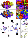
Structure of MVM VP2. The MVMpb structure is shown in red, that of MVMpe is in brown, and that of MVMi is in blue. (A to C) Electron density (2Fo-Fc) maps (gray wire) for MVMpb, MVMpe, and MVMi amino acids 316 to 322, respectively, containing MVMi/p differences at positions 317 and 321, the allotropic determinants. The MVMpb and MVMi structures are superimposed in panels A and C, and those of MVMpb and MVMpe are superimposed in panel B. (D and E) Electron density (2Fo-Fc) maps (gray wire) for MVMpb and MVMi amino acids 362 to 368, respectively, containing MVMi/p differences at positions 362, 366, and 368. (F and G) Electron density (2Fo-Fc) maps (gray wire) for MVMpb and MVMi amino acids 150 to 173, respectively, containing an MVMi/p difference at position 160. (H) Superimposition of coil representations for the amino acid stretch shown in panels F and G for MVMpb (red) and MVMi (blue). The orientations of the structures inpanels F to H are perpendicular to the icosahedral fivefold axis. (I and J) Coil representations for residues 157 to 164, with altered conformations in MVMpb and MVMi, respectively. The approximate fivefold axis is shown in the filled pentagon. (K) Superimposition of the ribbon diagrams of MVMpb (red) and MVMi (blue) VP2, illustrating β-strand, helical, and loop regions. Conserved β-strands βB to βI, helix αA, and residues 39 and 587, the first N-terminal residue modeled and the C-terminal residue, respectively, are labeled. The approximate icosahedral twofold (filled oval), threefold (filled triangle), and fivefold (filled pentagon) axes are shown. This figure was generated with the BOBSCRIPT (22) (A to J) and PyMol (21) (K) programs.
FIG. 2.
FIG. 2.
Depth-cued surface representations of the MVM capsid. (A) The surface topologies of MVMpb (red) and MVMi (blue) are shown with fivefold (5f), threefold (3f), and twofold (2f) axes labeled on the MVMpb capsid. A viral asymmetric unit is depicted by a triangle bounded by two threefold axes divided by a line drawn through the twofold axis, and a fivefold axis. (B to D) Close-up views of the MVMpb (red) and MVMi (blue) capsid surfaces at the fivefold (B), threefold (C), and twofold (D) icosahedral axes. The panel at the bottom depicts the color range (in Å) for the depth-cued distances from the viral center of the particles. This figure was generated by the GRASP program (42).
FIG. 3.
FIG. 3.
Structural clustering of MVMi/p amino acid differences. (A) (Right) Surface representation of the MVMpb capsid showing VP2 molecules related to a reference monomer (ref, in red) by icosahedral twofold (2f, in pink), threefold (3f1, in purple; 3f2, in yellow; and 3f3, in salmon), and fivefold (5f, in orange) icosahedral symmetry operations. (Left) Close-up view of the MVMpb icosahedral twofold axes, with the positions of surface MVMi/p amino acid differences colored and labeled: green for i/p differences (residues 232, 321, 362, 366, 368, 388, 410, and440; residue 317 is not visible in the view shown, and residue 551 is buried) and blue for forward fibrotropic mutations (residues 399, 553, and 558; residue 460 is buried under residue 399). (B) (Right) Surface representation of the MVMi capsid showing the ref (blue) and the 2f, 3f1, 3f2, 3f3, and 5f1 monomers (orange, pink, yellow, grey, and salmon, respectively). (Left) Close-up view of the MVMi icosahedral twofold axes, with the residues colored and labeled as in panel A, except that the forward fibrotropic residues appear red rather than blue. A viral asymmetric unit, as described in the legend to Fig. 2, is shown in panels A and B. White ovals represent approximate icosahedral twofold axes. (C) Side chain conformations of 12 of the 13 differing MVMi/p residues (residues 232, 317, 321, 362, 366, 368, 388, 402, 410, 440, 455, and 551) that are ordered within VP2 and are clustered from symmetry-related monomers. These are located at or surrounding the icosahedral twofold axes and on the shoulder of the threefold protrusions. (D to F) Close-up views of the intra- and intersubunit interactions involving allotropic and forward fibrotropic mutation residues. (D) Superimposition of the MVMpb (red) and MVMi (blue) residues 321, 368, 399, 400, and 460. (E and F) Amino acids for the MVMpb and MVMi capsids, respectively, colored according to atom type (yellow, carbon; red, oxygen; blue, nitrogen), with dashed lines labeled to indicate the distance between the charged atoms that could engage in ionic (H-bond) interactions. This figure was generated with the PyMol (21) (A and B) and BOBSCRIPT (22) (C to F) programs.
FIG. 4.
FIG. 4.
MVM capsid stability. Shown are the percentages of hemagglutination activity (y axis) in mouse erythrocytes for the MVMpb (open bars) MVMpe (hatched bars), and MVMi (solid bars) capsids after incubation (10 min) at the different temperatures (25 to 85°C, x axis) and the pHs indicated. Titers (percentage of HA normalized per microgram of capsid protein) are the reciprocal limiting dilutions causing hemagglutination. DL, detection limit. Error bars, standard errors of the means from three independent experimental runs.
FIG. 5.
FIG. 5.
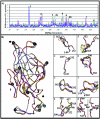
Comparison of the VP2 structures of CPV, FPV, PPV, and MVM. (A) Plot of the Cα differences between the refined VP2 structures of MVMpb and MVMi (blue), CPV (wt full capsids; PDB accession no. 4DPV) (grey), CPV-N93D (mutant empty capsids; PDB accession no. 1P5Y) (cyan), FPV (wt empty capsids; PDB accession no. 1C8E) (green), and PPV (baculovirus-expressed empty capsids; PDB accession no. 1K3V) (pink), calculated using the Homology program (49). Regions at a Cα difference of ≥2.0 Å between MVMpb and MVMi are labeled 1 to 8. This figure was generated with Excel as part of the Microsoft Office package. (B) Superimposition of a coil representation of the VP2 backbone atoms of MVMpb (red), MVMi (blue), CPV (wt and mutant structures; PDB accession no. 2CAS, 4DPV, 1P5Y, and 1P5W in yellow, grey, orange, and cyan, respectively), FPV (PDB accession no. 1C8E) (green), and PPV (PDB accession no. 1K3V) (pink). Variable regions equivalent to regions 1 to 8 in panel A are labeled. The icosahedral twofold, threefold, and fivefold axes are shown as the filled oval, triangle, and pentagon, respectively. Panels on the right show close-up views of the structures at variable regions 1 to 8. This figure was generated by PyMol (21).
FIG. 6.
FIG. 6.
Correlation of variable parvovirus capsid surface regions with tropism and pathogenicity determinants. (A) Variable regions 1 to 8 (highlighted in Fig. 5) were mapped onto MVMpb VP2 icosahedral symmetry-related monomers as colored balls and labeled. Colors: red, ref; pink, twofold (2f); light to dark green, threefold (3f1 to 3f3); cyan, fivefold (5f1 to 5f4). (B) The Cα positions of residues implicated in tropism and pathogenicity determination for MVM, CPV/FPV, PPV, and AMDV are shown as colored balls mapped to MVMpb VP2 icosahedral symmetry-related monomers. Ball colors: red, MVM allotropic residues (317 and 321); blue, MVMi forward mutations conferring fibrotropism (residues 399, 553, and 558); yellow, CPV tropism and pathogenicity determinants, and proposed receptor attachment residues (residues 93, 300, and 323); black, CPV sialic acid binding residues (residues 377, 396, and 397); green, FPV tropism and pathogenicity determinants (residues 80, 564, and 568); pink, PPV tropism and pathogenicity determinants (residues 377, 388, and 434); orange, AMDV tropism and pathogenicity determinants (residues 352, 395, 434, and 534). The viral asymmetry unit, as defined in Fig. 2A, is shown in both panels. Both panels are shown approximately down the icosahedral twofold axes. This figure was generated by the BOBSCRIPT program (22).

Similar articles

Cited by

References

    1. Agbandje, M., S. Kajigaya, R. McKenna, N. S. Young, and M. G. Rossmann. 1994. The structure of human parvovirus B19 at 8 Å resolution. Virology 203:106-115. - PubMed
    1. Agbandje, M., R. McKenna, M. G. Rossmann, M. L. Strassheim, and P. R. Parrish. 1993. Structure determination of feline panleukopenia virus empty particles. Proteins 16:155-171. - PubMed
    1. Agbandje-McKenna, M., A. L. Llamas-Saiz, F. Wang, P. Tattersall, and M. G. Rossmann. 1998. Functional implications of the structure of the murine parvovirus, minute virus of mice. Structure 6:1369-1381. - PubMed
    1. Agbandje-McKenna, M., C. R. Parrish, and M. G. Rossmann. 1995. The structure of parvoviruses. Semin. Virol. 6:299-309.
    1. Astell, C. R., E. M. Gardiner, and P. Tattersall. 1986. DNA sequence of the lymphotropic variant of minute virus of mice, MVM(i), and comparison with the DNA sequence of the fibrotropic prototype strain. J. Virol. 57:656-669. - PMC - PubMed

Publication types

Substances

LinkOut - more resources