. 2005 Sep;79(17):11082-94.
doi: 10.1128/JVI.79.17.11082-11094.2005.

The cellular TATA binding protein is required for rep-dependent replication of a minimal adeno-associated virus type 2 p5 element

Affiliations

The cellular TATA binding protein is required for rep-dependent replication of a minimal adeno-associated virus type 2 p5 element

Achille François et al. J Virol. 2005 Sep.

Abstract

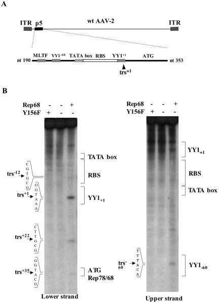
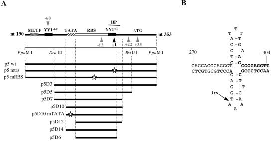
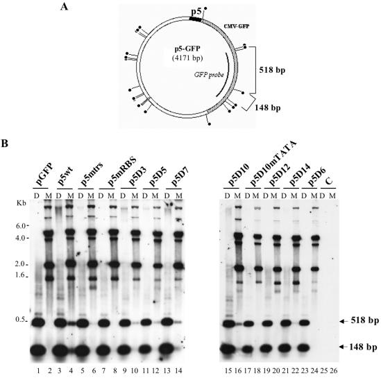
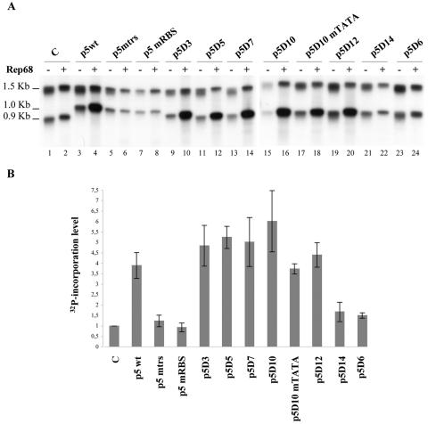
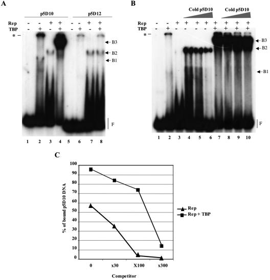
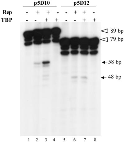
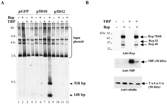
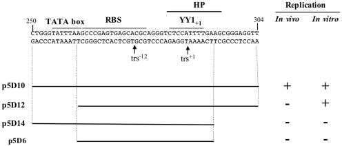
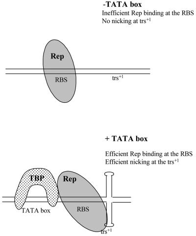
The p5 promoter region of adeno-associated virus type 2 (AAV-2) is a multifunctional element involved in rep gene expression, Rep-dependent replication, and site-specific integration. We initially characterized a 350-bp p5 region by its ability to behave like a cis-acting replication element in the presence of Rep proteins and adenoviral factors. The objective of this study was to define the minimal elements within the p5 region required for Rep-dependent replication. Assays performed in transfected cells (in vivo) indicated that the minimal p5 element was composed by a 55-bp sequence (nucleotides 250 to 304 of wild-type AAV-2) containing the TATA box, the Rep binding site, the terminal resolution site present at the transcription initiation site (trs(+1)), and a downstream 17-bp region that could potentially form a hairpin structure localizing the trs(+1) at the top of the loop. Interestingly, the TATA box was absolutely required for in vivo but dispensable for in vitro, i.e., cell-free, replication. We also demonstrated that Rep binding and nicking at the trs(+1) was enhanced in the presence of the cellular TATA binding protein, and that overexpression of this cellular factor increased in vivo replication of the minimal p5 element. Together, these studies identified the minimal replication origin present within the AAV-2 p5 promoter region and demonstrated for the first time the involvement of the TATA box, in cis, and of the TATA binding protein, in trans, for Rep-dependent replication of this viral element.


Figures

FIG. 1.
FIG. 1.
(A) Schematic view of the wild-type AAV-2 genome and of the wild-type p5 element. (B) Mapping of the Rep68 nicking sites on the p5 element. Nicking reactions were performed on a p5 substrate (nucleotides 190 to 353 of wild-type AAV-2) labeled on either strand in the presence of 1 pmol of Rep68 as described in the Materials and Methods section. The reaction products were resolved on 6% denaturing polyacrylamide gels along with a sequencing reaction to precisely map the nicking sites. The five identified nicking sites are indicated, as well as the position of the outstanding p5 elements.
FIG. 2.
FIG. 2.
(A) Presentation of the wild-type and mutated p5 elements. The arrowheads indicate the position of the Rep68 nicking sites (black: major cleavage site at trs+1; gray: other minor trs sites). The putative hairpin (HP) structure overlapping trs+1 is represented by a thick black line. White stars show the positions of the point mutations that were introduced within the p5 sequence (37). (B) Putative hairpin secondary structure present at the p5 transcription initiation site (trs+1). Bold nucleotides indicate the deletion of the hairpin structure present in mutants p5D14 and p5D6.
FIG. 3.
FIG. 3.
Identification of a minimal p5 element (p5D10) able to replicate in adenovirus-infected and Rep-expressing cells. (A) Map of the p5-GFP plasmid indicating the position of the DpnI/MboI sites and the GFP fragment used as a probe. (B) In vivo replication assay of the p5-containing plasmids. Total DNA extracted 48 h posttransfection of adenovirus type 5-infected HeRC32 cells, was digested with either DpnI (D) or MboI (M) and analyzed by Southern blot using a GFP probe. The two expected MboI or DpnI digestion products (518 and 148 bp) hybridizing to the GFP probe are indicated by an arrow. The upper bands visible in the MboI-digested samples represent input plasmid DNA. The amount of sample loaded in each lane was normalized using an internal control measuring the transfection efficiency (see Materials and Methods).
FIG. 4.
FIG. 4.
Identification of a minimal p5 (p5D12) element able to replicate in a cell-free assay. (A) In vitro replication assay of the p5-containing plasmids. Plasmids were digested with AflIII and XmnI and incubated with cell extracts from adenovirus type 5-infected HeLa cells (75 μg) and 300 ng of either Rep68 (+) or β-galactosidase (−) protein in the presence of [α-32P]dCTP. The reaction products were visualized by autoradiography of the membrane. The p5 element is contained in the lower DNA band. C, control pSP72 plasmid with no p5 element. (B) Quantification of the in vitro replication assays. The level of Rep-dependent replication of each individual p5-containing fragment was expressed as 32P incorporation calculated as described in Materials and Methods. The mean and standard deviation were calculated from three independent experiments.
FIG. 5.
FIG. 5.
Evidence for the formation of Rep and TBP complexes on the minimal p5D10 promoter element. (A) Enhanced binding of Rep68 to the p5D10 element in the presence of human TBP. EMSA of radiolabeled p5D10 (lanes 1 to 4) or p5D12 (lanes 5 to 8) DNA fragments incubated with either purified human TBP, Rep68, or both proteins. (B) Competition EMSA. The binding was performed using radiolabeled p5D10 DNA substrate incubated either alone or with the indicated proteins. The binding reactions were then completed with an excess (0-, 30-, 100-, or 300-fold) of unlabeled p5D10 competitor DNA and kept at 30°C for an additional 30 min. (C) Quantification of the amount of bound p5D10 labeled DNA. The amount of free DNA (F) in each lane was quantified by PhosphorImager analysis of the gel presented in panel B and the % of bound p5D10 DNA was calculated by subtracting each value from that obtained in the absence of any protein (lane 1). The asterisk in panels A and B indicates the position of the wells in the gel.
FIG. 6.
FIG. 6.
Effect of TBP on Rep endonuclease activity on the minimal p5D10 element. Nicking assays were conducted on either p5D10 or p5D12 DNA substrates in the presence (+) or absence (−) of purified TBP and Rep68. The final amount of recombinant protein in the reaction was kept constant by adding purified β-galactosidase. Reactions were performed in the same conditions as those described for EMSA except that they contained 1 mM ATP, 6.25 mM MgCl2 and that they were incubated for 1 h at 37°C after addition of Rep68. Reaction products were resolved on 8% denaturing polyacrylamide gels. The open arrowheads indicate the position of the uncut p5D10 and p5D12 fragments labeled at both termini whereas the solid arrows indicate the position of the product expected after nicking at the trs+1 (see Fig. 2).
FIG. 7.
FIG. 7.
Effect of TBP overexpression on in vivo replication of the minimal p5D10 element. (A) Southern blot analysis of in vivo replication products obtained by transfecting adenovirus type 5-infected 293 cells with plasmids pGFP, p5D10-GFP, and p5D12-GFP in the presence (+) or absence (−) of plasmids pCMV-rep (Rep) and pXJ41-TBP (TBP). Total DNA was digested with MboI and hybridized to a GFP probe. (B) Analysis of Rep and TBP expression in transfected cells. Adenovirus-infected 293 cells were transfected with plasmid p5D10-GFP and either plasmid pCMV-rep (Rep) or plasmid pXJ41-hTBP (TBP) or both. Total proteins were analyzed by Western blot using an anti-Rep antibody (303.9). The membrane was then stripped and reprobed with an anti-TBP antibody and then with an antitubulin antibody. Note that endogenous TBP was visible only after prolonged exposure of the blot.
FIG. 8.
FIG. 8.
Summary of the results using in vivo and in vitro replication assays performed with the minimal p5 elements (p5D10, p5D12, p5D14, and p5D6). +, replication; −, no replication.
FIG. 9.
FIG. 9.
Theoretical model for Rep-TBP interaction on the p5 element. See text for details.

References

    1. Berns, K. I., and C. Giraud. 1996. Biology of adeno-associated virus. Curr. Top. Microbiol. Immunol. 218:1-23. - PubMed
    1. Boyd, J. M., P. M. Loewenstein, Q.-Q. Tang, L. Yu, and M. Green. 2002. Adenovirus E1A N-terminal amino acid sequence requirements for repression of transcription in vitro and in vivo correlate with those required for E1A interference with TBP-TATA complex formation. J. Virol. 76:1461-1474. - PMC - PubMed
    1. Brister, J. R., and N. Muzyczka. 2000. Mechanism of Rep-mediated adeno-associated virus origin nicking. J. Virol. 74:7762-7771. - PMC - PubMed
    1. Brister, J. R., and N. Muzyczka. 1999. Rep-mediated nicking of the adeno-associated virus origin requires two biochemical activities, DNA helicase activity and transesterification. J. Virol. 73:9325-9336. - PMC - PubMed
    1. Burgess Hickman, A., D. R. Ronning, Z. N. Perez, R. M. Kotin, and F. Dyda. 2004. The nuclease domain of adeno-associated virus Rep coordinates replication initiation using two distinct DNA recognition interfaces. Mol. Cell 13:403-414. - PubMed

Publication types

Substances

LinkOut - more resources