. 2005 Sep;79(17):11095-104.
doi: 10.1128/JVI.79.17.11095-11104.2005.

Monoclonal antibody AP33 defines a broadly neutralizing epitope on the hepatitis C virus E2 envelope glycoprotein

Affiliations

Monoclonal antibody AP33 defines a broadly neutralizing epitope on the hepatitis C virus E2 envelope glycoprotein

Ania Owsianka et al. J Virol. 2005 Sep.

Abstract

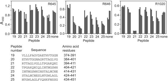
Hepatitis C virus (HCV) remains a significant threat to the general health of the world's population, and there is a pressing need for the development of new treatments and preventative vaccines. Here, we describe the generation of retrovirus-based pseudoparticles (HCVpp) incorporating a panel of full-length E1E2 clones representative of the major genotypes 1 through 6, and their application to assess the reactivity and neutralizing capability of antisera and monoclonal antibodies raised against portions of the HCV E2 envelope protein. Rabbit antisera raised against either the first hypervariable region or ectodomain of E2 showed limited and strain specific neutralization. By contrast, the monoclonal antibody (MAb) AP33 demonstrated potent neutralization of infectivity against HCVpp carrying E1E2 representative of all genotypes tested. The concentration of AP33 required to achieve 50% inhibition of infection by HCVpp of diverse genotypes ranged from 0.6 to 32 mug/ml. The epitope recognized by MAb AP33 is linear and highly conserved across different genotypes of HCV. Thus, identification of a broadly neutralizing antibody that recognizes a linear epitope is likely to be of significant benefit to future vaccine and therapeutic antibody development.


Figures

FIG. 1.
FIG. 1.
Analysis of E1E2 of diverse HCV genotypes. (A) A series of threefold dilutions of lysates of HEK 293T cells cotransfected with the MLV packaging/transfer vector and a plasmid expressing E1E2 of different HCV genotypes were analyzed for the presence of E2 by GNA capture ELISA using MAb AP33 (•) or ALP98 (○) (at 1 μg/ml) or the polyclonal antiserum R646 (▴) (1:10,000 dilution) as described in Materials and Methods. (B) Radiolabeled proteins in the lysate of HEK 293T cells cotransfected with different plasmids as described in A above were immunoprecipitated using a mixture of anti-E2 MAbs AP33 and ALP98. The immune complexes were analyzed by SDS-10% PAGE under reducing conditions. Positions of protein size markers are shown (in kDa).
FIG. 1.
FIG. 1.
Analysis of E1E2 of diverse HCV genotypes. (A) A series of threefold dilutions of lysates of HEK 293T cells cotransfected with the MLV packaging/transfer vector and a plasmid expressing E1E2 of different HCV genotypes were analyzed for the presence of E2 by GNA capture ELISA using MAb AP33 (•) or ALP98 (○) (at 1 μg/ml) or the polyclonal antiserum R646 (▴) (1:10,000 dilution) as described in Materials and Methods. (B) Radiolabeled proteins in the lysate of HEK 293T cells cotransfected with different plasmids as described in A above were immunoprecipitated using a mixture of anti-E2 MAbs AP33 and ALP98. The immune complexes were analyzed by SDS-10% PAGE under reducing conditions. Positions of protein size markers are shown (in kDa).
FIG. 2.
FIG. 2.
Neutralization of HCVpp infection by anti-E2 antibodies. (A) HCVpp harboring the genotype 1a H77 E1E2 were preincubated with 1:120 dilutions of anti-E2 sera R645, R646, R1020, and R1021 along with their preimmune (PI) counterparts or 50 μg/ml MAb AP33 prior to infection of Huh-7 cells. (B) Neutralization assay performed following incubation of HCVpp derived from constructs expressing E1E2 of genotypes 1a (strain H77), 1b (UKN1B12.16), 2a (UKN2A2.4), or 2b (UKN2B1.1) with 1:100 dilutions of polyclonal sera and 50 μg/ml MAb AP33. (C) Neutralization of HCVpp derived from E1E2 of three different genotype 1a subtypes (strains H77, UKN1A14.8, and UKN1A14.36) using a range of R646 concentrations.
FIG. 3.
FIG. 3.
Neutralization by MAb AP33 of HCVpp derived from diverse genotypes. HCVpp were preincubated for 1 h at 37°C with different concentrations of purified MAb AP33 prior to infection of Huh-7 cells. The neutralizing activity of the antibody is expressed as percentage of inhibition of the infectious titers.
FIG. 4.
FIG. 4.
Epitope mapping of anti-E2 antibodies in sera by peptide competition ELISA. The sera R1020 (1:1,000), R645 (1:1,000), and R646 (1:10,000) were preincubated with 31 μg/ml (first bar), 93 μg/ml (second bar), 280 μg/ml (third bar), and 830 μg/ml (fourth bar) of a series of overlapping peptides for 1 h at 37°C. The antibody/peptide mix was then tested for reactivity against soluble E2 in a GNA capture ELISA. The details of the peptides used are shown at the bottom.
FIG. 5.
FIG. 5.
R646 antiserum contains antibodies recognizing both conformational as well as linear epitopes in E2. Approximately 15 μg of purified R646 IgGs were either left untreated (solid circles), preadsorbed with GNA (open circles), native E2 on GNA (open triangles), or denatured E2 on GNA (solid triangle) and tested at different dilutions (A) for recognition of native E2 in a GNA capture ELISA and (B) for neutralization of HCVpp generated from genotype 1a H77 E1E2.
FIG. 6.
FIG. 6.
Alignment of functional proteins of diverse genotypes, covering the regions possessing epitopes recognized by anti-E2 antibodies R646 and AP33 (A) and ALP98 (B). The MAb AP33 and ALP98 epitopes are highlighted in bold, and regions reactive with R646 antiserum are underscored. The sequence of strain H77 was published by Yanagi et al. (65). The accession numbers of our sequences have been deposited with GenBank. UKN1A14.36, AY799303; UKN1B12.16, AY734974; UKN2A2.4, AY734979; UKN2B1.1, AY734981; UKN3A13.6, AY894683; UKN4.21.16, AY734987; UKN5.14.4, AY785283; UKN5.15.11, AY894682; UKN6.5.340, AY736194.

References

    1. Allander, T., K. Drakenberg, A. Beyene, D. Rosa, S. Abrignani, M. Houghton, A. Widell, L. Grillner, and M. A. A. Persson. 2000. Recombinant human monoclonal antibodies against different conformational epitopes of the E2 envelope glycoprotein of hepatitis C virus that inhibit its interaction with CD81. J. Gen. Virol. 81:2451-2459. - PubMed
    1. Alter, M. J., H. S. Margolis, K. Krawczynski, F. N. Judson, A. Mares, W. J. Alexander, H. Pin Ya, J. K. Miller, M. A. Gerber, R. E. Sampliner, E. L. Meeks, and M. J. Beach. 1992. The natural history of community-acquired hepatitis C in the United States. N. Engl. J. Med. 327:1899-1905. - PubMed
    1. Andre, P., F. Komurian-Pradel, S. Deforges, M. Perret, J. L. Berland, M. Sodoyer, S. Pol, C. Brechot, G. Paranhos-Baccala, and V. Lotteau. 2002. Characterization of low- and very-low-density hepatitis C virus RNA-containing particles. J. Virol. 76:6919-6928. - PMC - PubMed
    1. Anonymous. 1999. Global surveillance and control of hepatitis C. J. Viral Hepat. 6:35-47. - PubMed
    1. Barth, H., C. Schafer, M. I. Adah, F. Zhang, R. J. Linhardt, H. Toyoda, A. Kinoshita-Toyoda, T. Toida, T. H. Van Kuppevelt, E. Depla, F. Von Weizsacker, H. E. Blum, and T. F. Baumert. 2003. Cellular binding of hepatitis C virus envelope glycoprotein E2 requires cell surface heparan sulfate. J. Biol. Chem. 278:41003-41012. - PubMed

Publication types

LinkOut - more resources