. 2005 Sep;79(17):11269-79.
doi: 10.1128/JVI.79.17.11269-11279.2005.

Are ducks contributing to the endemicity of highly pathogenic H5N1 influenza virus in Asia?

Affiliations

Are ducks contributing to the endemicity of highly pathogenic H5N1 influenza virus in Asia?

K M Sturm-Ramirez et al. J Virol. 2005 Sep.

Abstract

Wild waterfowl are the natural reservoir of all influenza A viruses, and these viruses are usually nonpathogenic in these birds. However, since late 2002, H5N1 outbreaks in Asia have resulted in mortality among waterfowl in recreational parks, domestic flocks, and wild migratory birds. The evolutionary stasis between influenza virus and its natural host may have been disrupted, prompting us to ask whether waterfowl are resistant to H5N1 influenza virus disease and whether they can still act as a reservoir for these viruses. To better understand the biology of H5N1 viruses in ducks and attempt to answer this question, we inoculated juvenile mallards with 23 different H5N1 influenza viruses isolated in Asia between 2003 and 2004. All virus isolates replicated efficiently in inoculated ducks, and 22 were transmitted to susceptible contacts. Viruses replicated to higher levels in the trachea than in the cloaca of both inoculated and contact birds, suggesting that the digestive tract is not the main site of H5N1 influenza virus replication in ducks and that the fecal-oral route may no longer be the main transmission path. The virus isolates' pathogenicities varied from completely nonpathogenic to highly lethal and were positively correlated with tracheal virus titers. Nevertheless, the eight virus isolates that were nonpathogenic in ducks replicated and transmitted efficiently to naïve contacts, suggesting that highly pathogenic H5N1 viruses causing minimal signs of disease in ducks can propagate silently and efficiently among domestic and wild ducks in Asia and that they represent a serious threat to human and veterinary public health.


Figures
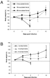
FIG. 1.
FIG. 1.
Weight loss of mallards infected with H5N1 influenza viruses isolates. Inoculated ducks were infected with 106 to 107 EID50 of virus and housed with contact ducks within 1 hour of inoculation, sharing food and water. Ducks were weighed daily for 10 days. Virus isolates were grouped based on their pathogenic potential in ducks as causing no mortality among either inoculated or contact ducks (I), causing death among inoculated ducks only (II), and causing death in both inoculated and contact ducks (III). Data points and error bars represent the averages and ranges of weight loss caused by virus isolates in each pathogenic group in inoculated ducks (A) and contact ducks (B).
FIG. 2.
FIG. 2.
Survival curve of mallards infected with different H5N1 influenza virus isolates. Ducks were observed for 14 days after inoculation with 106 to 107 EID50 of virus via the natural route. Ducks that exhibited severe neurological signs were euthanized, and their deaths were recorded on the following day of observation. Although survival curves were generated for all 23 virus isolates, for clarity only representative curves of a few isolates per pathogenicity group were included on the graph. Classification is as follows: low pathogenicity, A/Thai/1(Kan-1)/04 and A/Ck/PP/BPPV3/04; high pathogenicity, A/Dk/VN/40D/04, A/Ck/VN/48C/04, A/Dk/Thai/71.1/04, and A/VN/1203/04.
FIG. 3.
FIG. 3.
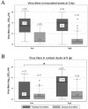
Tracheal and cloacal virus titers in mallards infected with H5N1 influenza viruses. Inoculated ducks were infected with 106 to 108.5 EID50 of virus and then housed with contact ducks within 1 hour of inoculation, sharing food and water. Tracheal and cloacal swabs were collected at 3 and 5 days postinoculation and tested for the presence of influenza virus, and positive samples were subjected to titer determinations for infectivity by calculating the EID50. The data represented in this figure comprise virus titer data from this study and data from previously published work (24). The study population comprised 135 mallards: 70 experimentally inoculated ducks and 65 contact ducks infected with 35 different H5N1 virus isolates obtained in Asia from 1997 to 2004. Virus isolates were classified in two groups based on their pathogenic potential in ducks: low pathogenicity and high pathogenicity. Box plots represent the distribution of tracheal and cloacal virus titers in each pathogenic group (low and high) in inoculated ducks on day 3 postinfection (A) and in contact ducks on day 5 postinfection (B). Each box shows the median value of the data set (black line and associated numerical value), the interquartile range (upper and lower boundaries of the box), the 10th percentile and 90th percentile (lower and upper error bars, respectively), and the size of each group (n). *, P < 0.05 (Kruskal-Wallis equality-of-population test).
FIG. 4.
FIG. 4.
Tracheal and cloacal virus titers in mallards infected with H5N1 influenza viruses. Inoculated ducks were infected with 106 to 108.5 EID50 of virus and then housed with contact ducks within 1 hour of inoculation, sharing food and water. Tracheal and cloacal swabs were collected at 3 and 5 days postinoculation and tested for the presence of influenza virus, and positive samples were subjected to titer determinations for infectivity by calculating the EID50. The data represented in this figure comprise virus titer data from this study and data from previously published work (24). The study population comprised 135 mallards: 70 experimentally inoculated ducks and 65 contact ducks infected with 35 different H5N1 virus isolates obtained in Asia from 1997 to 2004. Virus isolates were classified in two groups based on their date of isolation (1997 to 2001 and 2002 to 2004). Box plots represent the distribution of tracheal (A) and cloacal (B) virus titers in inoculated and contact ducks at 3 and 5 dpi, respectively. Each box shows the median value of the data set (black line and associated numerical value), the interquartile range (upper and lower boundaries of the box), the 10th percentile and 90th percentile (lower and upper error bars, respectively), and the size of each group (n). *, significant difference in median value (P < 0.05; K-sample test on equality of median).

References

    1. Alexander, D. J., G. Parsons, and R. J. Manvell. 1986. Experimental assessment of the pathogenicity of eight avian influenza A viruses of H5 subtype for chickens, turkeys, ducks and quail. Avian Pathol. 15:647-662. - PubMed
    1. Capua, I., and F. Mutinelli. 2001. Mortality in Muscovy ducks (Cairina moschata) and domestic geese (Anser anser var. domestica) associated with natural infection with a highly pathogenic avian influenza virus of H7N1 subtype. Avian Pathol. 30:179-183. - PubMed
    1. Cauthen, A. N., D. E. Swayne, S. Schultz-Cherry, M. L. Perdue, and D. L. Suarez. 2000. Continued circulation in China of highly pathogenic avian influenza viruses encoding the hemagglutinin gene associated with the 1997 H5N1 outbreak in poultry and humans. J. Virol. 74:6592-6599. - PMC - PubMed
    1. Chen, H., G. Deng, Z. Li, G. Tian, Y. Li, P. Jiao, L. Zhang, Z. Liu, R. G. Webster, and K. Yu. 2004. The evolution of H5N1 influenza viruses in ducks in southern China. Proc. Natl. Acad. Sci. USA 101:10452-10457. - PMC - PubMed
    1. Claas, E. C., A. D. Osterhaus, R. van Beek, J. C. de Jong, G. F. Rimmelzwaan, D. A. Senne, S. Krauss, K. F. Shortridge, and R. G. Webster. 1998. Human influenza A H5N1 virus related to a highly pathogenic avian influenza virus. Lancet 351:472-477. - PubMed

Publication types