. 2005 Sep;79(17):11335-42.
doi: 10.1128/JVI.79.17.11335-11342.2005.

A severe acute respiratory syndrome-associated coronavirus-specific protein enhances virulence of an attenuated murine coronavirus

Affiliations

A severe acute respiratory syndrome-associated coronavirus-specific protein enhances virulence of an attenuated murine coronavirus

Lecia Pewe et al. J Virol. 2005 Sep.

Abstract

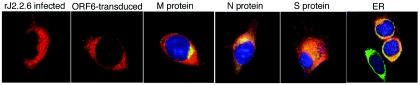
Most animal species that can be infected with the severe acute respiratory syndrome-associated coronavirus (SARS-CoV) do not reproducibly develop clinical disease, hindering studies of pathogenesis. To develop an alternative system for the study of SARS-CoV, we introduced individual SARS-CoV genes (open reading frames [ORFs]) into the genome of an attenuated murine coronavirus. One protein, the product of SARS-CoV ORF6, converted a sublethal infection to a uniformly lethal encephalitis and enhanced virus growth in tissue culture cells, indicating that SARS-CoV proteins function in the context of a heterologous coronavirus infection. Furthermore, these results suggest that the attenuated murine coronavirus lacks a virulence gene residing in SARS-CoV. Recombinant murine coronaviruses cause a reproducible and well-characterized clinical disease, offer virtually no risk to laboratory personnel, and should be useful for elucidating the role of SARS-CoV nonstructural proteins in viral replication and pathogenesis.


Figures

FIG. 1.
FIG. 1.
Expression of SARS-CoV nonstructural ORFs in rJ2.2-infected cells. (A) Genome of SARS-CoV, with ORFs and structural proteins (gray). (B) SARS-CoV nonstructural proteins were introduced into rJ2.2 by targeted recombination (shown for rJ2.2.6) as described in Materials Methods. As controls, a termination codon was introduced at residue 27 in rJ.2.2.6trunc and rJ2.2.6KO. The initiator methionine was also mutated in rJ2.2.6KO. Each construct was C terminus tagged with the influenza virus HA epitope. (C) The products of ORF3a, -6, and -7b were detected by Western blot analysis using anti-HA antibody. Lanes: 1, rJ2.2; 2, rJ2.2.6; 3, rJ2.2.3a; 4, rJ2.2.3b; 5, rJ2.2.7a; 6, rJ2.2.7b; 7, rJ2.2.8. (D) All of the inserted proteins were detected by IFA using anti-HA antibody. The products of ORF3b, -7a, and -8 are shown. Note that protein 3b was localized to the nucleus. No staining was detected in cells infected with rJ2.2 (WT).
FIG. 2.
FIG. 2.
Mortality, morbidity, and virus titers in mice infected with rJ2.2.6, rJ2.2.6trunc, or rJ2.2.6KO. Mice were infected with rJ2.2 (triangles), rJ2.2.6 (open circles), or rJ2.2.6trunc or rJ2.2.6KO (squares) and monitored for mortality (A), clinical disease (B), and weight loss (C). Groups of 6 rJ2.2-, 38 rJ2.2.6-, and 30 rJ2.2.6trunc- or rJ2.2.6KO-infected mice were used in these studies. (D) CNS virus titers were also determined. A total of 212 mice were used for this purpose. Significant increases in virus titers were detected in the CNS of rJ2.2.6-infected mice compared to mice infected with rJ2.2.6trunc+KO (day 1.5, P = 0.38; day 3, P = 0.23; day 6, P < 0.01; day 7, P = 0.056; day 9, P < 0.001). Significant increases in virus titers were detected in the CNS of rJ2.2.6-infected mice compared to mice infected with rJ2.2 (day 7, P < 0.05; day 9, P < 0.005). Data for days 0 to 9 p.i. are shown in panels B, C, and D for the rJ2.2.6-infected mice because only 23% of mice survived past this time. The data for rJ2.2.6trunc- and rJ2.2.6KO-infected mice were combined, since mice infected with these viruses developed the same clinical disease. (E) RNA was harvested from the brains of mice infected with rJ2.2.6 (open bars; n = 3) or rJ2.2.6KO (closed bars; n = 3) at each time point. The amount of N gene-specific RNA was quantified by real-time RT-PCR as described in Materials and Methods.
FIG. 3.
FIG. 3.
Virus titers and interferon responses in rJ2.2.6- and rJ2.2.6KO-infected L929 cells. (A) L929 cells were infected with rJ2.2.6 or rJ2.2.6KO at an MOI of 1. Cells were harvested in triplicate at the indicated times, and virus titers were measured on HeLa-MHVR cells. Open circles, rJ2.2.6; squares, rJ2.2.6KO. Titers and SEM are shown. (B) L929 cells infected with rJ2.2.6 (lanes 2, 4, and 6) or rJ2.2.6KO (lanes 3, 5, and 7) were pulse-labeled with Tran35S-label for 30 min at 17 h (lanes 2 and 3), 22 h (lanes 4 and 5), and 29 h (lanes 6 and 7) and processed as described in Materials and Methods. Lane 1 shows mock-infected cells. The N and S proteins are indicated. Fifty micrograms of protein was loaded per lane. (C) RNA was harvested at 16 h p.i. from L929 cells infected with rJ2.2.6 (lanes 1 and 2; two isolates), rJ2.2.6KO (lanes 3 and 4; two isolates), or rJ2.2 (lane 5) or mock-infected cells (lane 6) and analyzed as described in Materials and Methods. Ten micrograms of RNA was loaded per lane. Viral RNAs 1 (genomic) and 2 to 7 (subgenomic) are indicated on the right. (D) At 24 h prior to infection with rJ2.2 (dashed line), rJ2.2.6 (open circles), or rJ2.2.6KO (squares), cells in triplicate were treated with the indicated concentrations of IFN-β. After infection (MOI = 0.1), cells were then incubated for 20 h in the presence of the same concentration of IFN. Samples were then harvested and virus titers were determined on HeLa-MHVR cells.
FIG. 4.
FIG. 4.
Distribution of S and ORF6-HA proteins in infected cell cultures. 17Cl-1 cells infected with rJ2.2.6 (MOI = 0.05) were detergent solubilized at 24 h p.i. (A) The indicated number of cell equivalents were subjected to electrophoresis and immunoblotting using MAb 10G (kindly provided by F. Taguchi, National Institute of Neuroscience, Tokyo, Japan) and anti-HA MAb HA.11 (Covance, Inc.) to detect S and ORF6-HA, respectively. (B) Virions were concentrated from clarified media by pelleting through sucrose cushions as described in Materials and Methods, and proteins were similarly processed to reveal the proteins. S proteins were distributed in a ∼40:1 cell/virion ratio, while ORF6-HA proteins were distributed in a >300:1 cell/virion ratio.
FIG. 5.
FIG. 5.
Localization of ORF6-HA with MHV structural proteins and BiP. L929 cells were infected with rJ2.2.6 for 12 to 16 h. After fixation, cells were stained for HA and the MHV M, N, and S proteins or the endoplasmic reticulum (ER) protein BiP. L929 cells transduced with a retrovirus expressing ORF6-HA are also shown. The last four panels are merged to show colocalization of ORF6-HA with the indicated protein.
FIG. 6.
FIG. 6.
Partitioning of MHV N, M, and ORF6-HA proteins into hydrophobic and hydrophilic environments. 17Cl-1 cells infected with rJ.2.2.6 were collected at 16 h p.i., and proteins were partitioned into hydrophobic (Triton X-114 detergent) and hydrophilic (aqueous) phases, using M-PER reagents (Pierce Company, Rockford, IL). Proteins from the indicated cell equivalents were then subjected to sodium dodecyl sulfate-polyacrylamide gel electrophoresis, transferred to polyvinylidene difluoride membranes, and probed simultaneously with anti-N MAb 5A5.2 and Covance anti-HA MAb 1.1 (top panel). The blot was then stripped and reprobed with anti-M MAb 4B6.2 (bottom panel).

Similar articles

Cited by

References

    1. Bergmann, C. C., Q. Yao, M. Lin, and S. A. Stohlman. 1996. The JHM strain of mouse hepatitis virus induces a spike protein-specific Db-restricted CTL response. J. Gen. Virol. 77:315-325. - PubMed
    1. Castro, R. F., and S. Perlman. 1995. CD8+ T-cell epitopes within the surface glycoprotein of a neurotropic coronavirus and correlation with pathogenicity. J. Virol. 69:8127-8131. - PMC - PubMed
    1. Chinese SARS Molecular Epidemiology Consortium. 2004. Molecular evolution of the SARS coronavirus during the course of the SARS epidemic in China. Science 303:1666-1669. - PubMed
    1. Cinatl, J., B. Morgenstern, G. Bauer, P. Chandra, H. Rabenau, and H. W. Doerr. 2003. Treatment of SARS with human interferons. Lancet 362:293-294. - PMC - PubMed
    1. Dandekar, A. A., and S. Perlman. 2002. Virus-induced demyelination in nude mice is mediated by gamma delta T cells. Am. J. Pathol. 161:1255-1263. - PMC - PubMed

Publication types

MeSH terms

Substances

LinkOut - more resources