Comparative Study
. 2005 Sep;79(17):11366-81.
doi: 10.1128/JVI.79.17.11366-11381.2005.

Human immunodeficiency virus type 1 Vpr-dependent cell cycle arrest through a mitogen-activated protein kinase signal transduction pathway

Affiliations
Comparative Study

Human immunodeficiency virus type 1 Vpr-dependent cell cycle arrest through a mitogen-activated protein kinase signal transduction pathway

Naoto Yoshizuka et al. J Virol. 2005 Sep.

Abstract

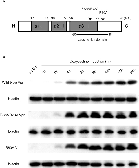
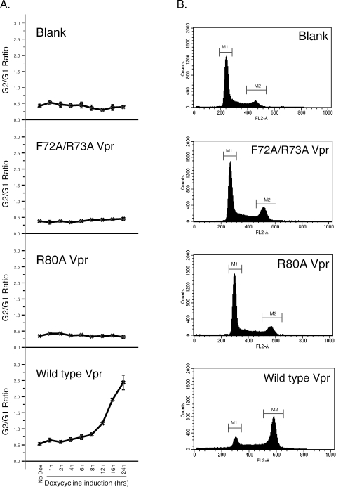
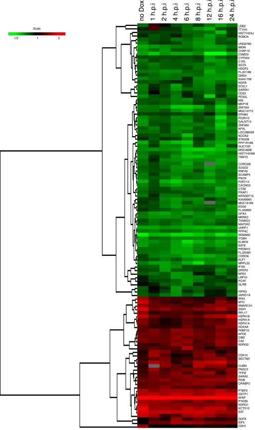
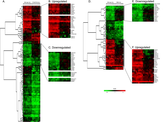
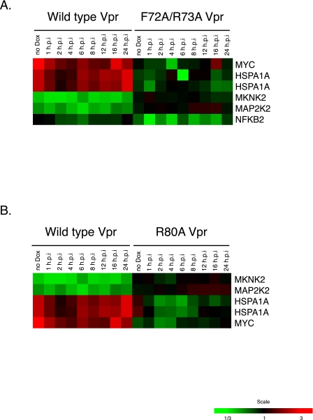
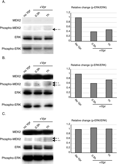
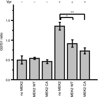
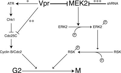
The human immunodeficiency virus type 1 (HIV-1) Vpr protein has important functions in advancing HIV pathogenesis via several effects on the host cell. Vpr mediates nuclear import of the preintegration complex, induces host cell apoptosis, and inhibits cell cycle progression at G(2), which increases HIV gene expression. Some of Vpr's activities have been well described, but some functions, such as cell cycle arrest, are not yet completely characterized, although components of the ATR DNA damage repair pathway and the Cdc25C and Cdc2 cell cycle control mechanisms clearly play important roles. We investigated the mechanisms underlying Vpr-mediated cell cycle arrest by examining global cellular gene expression profiles in cell lines that inducibly express wild-type and mutant Vpr proteins. We found that Vpr expression is associated with the down-regulation of genes in the MEK2-ERK pathway and with decreased phosphorylation of the MEK2 effector protein ERK. Exogenous provision of excess MEK2 reverses the cell cycle arrest associated with Vpr, confirming the involvement of the MEK2-ERK pathway in Vpr-mediated cell cycle arrest. Vpr therefore appears to arrest the cell cycle at G(2)/M through two different mechanisms, the ATR mechanism and a newly described MEK2 mechanism. This redundancy suggests that Vpr-mediated cell cycle arrest is important for HIV replication and pathogenesis. Our findings additionally reinforce the idea that HIV can optimize the host cell environment for viral replication.


Figures

FIG. 1.
FIG. 1.
Construction and analysis of the Vpr-expressing cell lines. (A) Diagram of the primary structure of wild-type Vpr representing α-helical domains I (a1-H), II (a2-H), and III (a3-H) and the leucine-rich domain. Mutation sites (F72A/R73A and R80A) are indicated by arrows. (B) Protein immunoblot analysis of Vpr protein expression regulated by doxycycline. Protein lysate was prepared from cells not treated with doxycycline (no Dox) and at 1, 2, 4, 6, 8, 12, 16, and 24 hours after doxycycline addition. Mouse anti-FLAG M2 monoclonal antibody was used to detect FLAG-tagged Vpr protein expression. As an internal control, mouse anti-human β-actin (b-actin) monoclonal antibody was used. a.a., amino acids.
FIG. 2.
FIG. 2.
Effects of induced wild-type and mutant Vpr expression on the cell cycle. (A) Kinetic analyses of G2/M cell cycle arrest after doxycycline treatment. Vpr-induced G2/M cell cycle arrest was measured by flow cytometry after DNA was stained with propidium iodide. The extent of G2/M arrest was evaluated by the G2/G1 ratio. The results represent the mean of at least three experiments. The standard deviations of the values are shown as error bars. (B) Representative data for Vpr-induced G2/M cell cycle arrest measured by flow cytometry at 24 h after doxycycline addition. The y axis represents the cell count, and the x axis represents the DNA content. The experiments were conducted at least three times.
FIG. 3.
FIG. 3.
Hierarchical clustering of differentially expressed cellular genes altered by the inducible expression of wild-type Vpr protein. The figure shows the hierarchical clustering of the cellular genes that showed statistically significant differences in expression (P < 0.001) and twofold changes in the expression of a cellular gene at least at one time point. Genes shown in red showed up-regulation, and those in green were down-regulated, while those that did not show any change with respect to a normalized matched control are shown in black (see color scale). The gray areas indicate missing data.
FIG. 4.
FIG. 4.
Differential expression of cellular genes following induction of wild-type and mutant Vpr expression. Comparison of differentially expressed cellular genes altered after Vpr expression between cells expressing (A) wild-type Vpr and F72A/R73A Vpr and (D) wild-type Vpr and R80A Vpr. Black bars on the right side of panels A and D indicate the clusters, which showed wild-type-specific differential regulation of gene expression. These clusters are highlighted in larger images with gene symbols. (B) Up- and (C) down-regulated in a comparison with F72A/R73A Vpr-expressing cells. (E) Down- and (F) up-regulated in a comparison with R80A Vpr-expressing cells. The genes shown in this figure passed two filtering criteria: (i) statistical significance (P < 0.001) and (ii) twofold changes in the expression of a cellular gene at least at one time point.
FIG. 5.
FIG. 5.
Differentially regulated genes related to the MAPK pathway. Assignment of these genes was performed with the Cancer Genome Anatomy Project pathway databases (http://cgap.nci.nih.gov/). Pathway information was provided by the Kyoto Encyclopedia of Genes and Genomes (http://www.genome.jp/kegg/).
FIG. 6.
FIG. 6.
Analysis of ERK2 phosphorylation status following induced expression of Vpr and abrogation of the effect by MEK2. Flp-In TREx 293 cells stably transfected with wild-type Vpr were transiently transfected with either a blank vector as a control plasmid (A), wild-type MEK2 (MEK2 WT) (B), or constitutively active mutant MEK2 (MEK2 CA) (C) and treated with 1 μg/ml doxycycline for the times indicated. The cells were lysed and analyzed for ERK phosphorylation using an anti-phospho-p44 (ERK1)/phospho-p42 (ERK2) phosphospecific antibody. Endogenous ERK1/2 expression levels were detected by immunoblotting using anti-ERK1/2 antibody. The higher band of the doublet detected by anti-ERK1/2 antibody is p44 (ERK1), and the lower band is p42 (ERK2). The signal due to phospho-ERK protein was quantitated using Image J software (http://rsb.info.nih.gov/ij/) and normalized to the signal due to endogenous ERK protein (p-ERK/ERK ratio). The relative changes in the p-ERK/ERK ratio (y axis) at each time point (+Vpr) were calculated by comparing the p-ERK/ERK ratio at each time point to the p-ERK/ERK ratio without doxycycline treatment (no Vpr). Endogenous or exogenous MEK2 expression levels were detected by immunoblotting using anti-MEK2 antibody. The phosphorylated forms of MEK1/2 were detected by immunoblotting using anti-phospho-MEK1/2 antibodies. * indicates the band for the phosphorylated form of MEK1/2, while ** indicates a cross-reacting band (1). We repeated the experiments at least three times. Representative data are shown.
FIG. 7.
FIG. 7.
Inhibition of Vpr-induced G2/M arrest by wild-type MEK2 and a constitutively active MEK2 mutant. Flp-In TREx 293 cells stably transfected with wild-type Vpr were transiently transfected with either a blank vector as a control plasmid, a wild-type MEK2 construct (MEK2 WT), or a constitutively active mutant of MEK2 (MEK2 CA). Twenty-four hours after transfection, the cells were treated with 1 μg/ml doxycycline for 24 h to induce Vpr (Vpr +) or left untreated (Vpr -). The results represent the mean of four experiments. The standard deviations of the values are shown as error bars. Differences between groups were examined for statistical significance using the unpaired t test. *, P < 0.02; **, P < 0.005.
FIG. 8.
FIG. 8.
Schematic representation of the putative signaling cascades mediating G2/M cell cycle arrest induced by Vpr protein. Previously described pathways, including those described by Zimmerman et al. (*) (46), who reported the involvement of the ATR-Chk1-mediated checkpoint pathway; Goh et al. (**) (16), who reported the direct inhibition of Cdc25C by Vpr; and Ussar and Voss (***) (38), who reported that the MEK2 knockdown results in centrosome amplification and multipolar spindle formation through reduced phosphorylation of RSK, are indicated. In our model, blockade of MEK2 activity also leads to cell cycle arrest at the G2/M checkpoint by decreasing ERK phosphorylation and abolishing the ability of the cells to recover from the G2/M checkpoint arrest.

Similar articles

Cited by

References

    1. Abbott, D. W., and J. T. Holt. 1999. Mitogen-activated protein kinase kinase 2 activation is essential for progression through the G2/M checkpoint arrest in cells exposed to ionizing radiation. J. Biol. Chem. 274:2732-2742. - PubMed
    1. Chowdhury, I. H., X. F. Wang, N. R. Landau, M. L. Robb, V. R. Polonis, D. L. Birx, and J. H. Kim. 2003. HIV-1 Vpr activates cell cycle inhibitor p21/Waf1/Cip1: a potential mechanism of G2/M cell cycle arrest. Virology 305:371-377. - PubMed
    1. Cohen, E. A., G. Dehni, J. G. Sodroski, and W. A. Haseltine. 1990. Human immunodeficiency virus vpr product is a virion-associated regulatory protein. J. Virol. 64:3097-3099. - PMC - PubMed
    1. Cohen, E. A., E. F. Terwilliger, Y. Jalinoos, J. Proulx, J. G. Sodroski, and W. A. Haseltine. 1990. Identification of HIV-1 vpr product and function. J. Acquir. Immune Defic. Syndr. 3:11-18. - PubMed
    1. Connor, R. I., B. K. Chen, S. Choe, and N. R. Landau. 1995. Vpr is required for efficient replication of human immunodeficiency virus type-1 in mononuclear phagocytes. Virology 206:935-944. - PubMed

Publication types

Substances