. 2005 Aug 15;19(16):1885-93.
doi: 10.1101/gad.1333905.

Methylation of histone H4 by arginine methyltransferase PRMT1 is essential in vivo for many subsequent histone modifications

Affiliations

Methylation of histone H4 by arginine methyltransferase PRMT1 is essential in vivo for many subsequent histone modifications

Suming Huang et al. Genes Dev. .

Abstract

PRMT1 is a histone methyltransferase that methylates Arg3 on histone H4. When we used siRNA to knock down PRMT1 in an erythroid cell line, it resulted in nearly complete loss of H4 Arg3 methylation across the chicken beta-globin domain, which we use as a model system for studying the relationship of gene activity to histone modification. We observed furthermore a domain-wide loss of histone acetylation on both histones H3 and H4, as well as an increase in H3 Lys9 and Lys27 methylation, both marks associated with inactive chromatin. To determine whether the effect on acetylation was directly related to the loss of H4 Arg3 methylation, we performed an in vitro acetylation reaction on chromatin isolated from PRMT1-depleted cells. We found that nucleosomes purified from these cells, and depleted in methylation at Arg3, are readily acetylated by nuclear extracts from the same cells, if and only if the nucleosomes are incubated with PRMT1 beforehand. Thus, methylation of histones by PRMT1 was sufficient to permit subsequent acetylation. Consistent with earlier reports of experiments in vitro, H4 Arg3 methylation by PRMT1 appears to be essential in vivo for the establishment or maintenance of a wide range of "active" chromatin modifications.


Figures
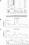
Figure 1.
Figure 1.
Pattern of histone H4 methylation at Arg3 across the β-globin locus at different erythroid development stages. (A) Map of the chicken β-globin locus. The folate receptor gene is located at the 5′-end of the locus. HSA is a hypersensitive site associated with FR expression. A second hypersensitive site, HS4, located 5′ upstream of the open globin domain, marks the insulator element of the β-globin. A 16-kb condensed chromatin segment is located between hypersensitive sites HSA and HS4. Downstream of HS4, there are three hypersensitive sites, HS3 to HS1, comprising part of the globin locus control region. βA/ε is a strong enhancer lying between the βA- and βε-globin genes. 3′HS marks the 3′-end of the β-globin locus and possesses enhancer-blocking properties only. The name and location of primer pairs and Taqman probes used for the ChIP analy sis are shown below the map. Above the map are data for four chicken cell types representing different development stages: 6C2 is arrested in the CFU-E (colony-forming unit-erythroid) stage, and 10-d erythrocytes are taken from the embryonic circulation. Expression of the individual genes is indicated by + and –. Y indicates the presence of the hypersensitive sites. (B,C) Pattern of H4 Arg3 methylation across the chicken β-globin neighborhood. Results of ChIP with antibody specific to dimethyl Arg3 on histone H4; data from 6C2 and 10-d chicken embryonic erythrocytes. Chromatin preparation and IP procedures are described in Materials and Methods and in Litt et al. (2001b).
Figure 2.
Figure 2.
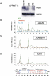
Both histone H3 and H4 acetylation are dependent on PRMT1 activity. (A) siRNA-mediated knock-down of PRMT1 expression in 6C2 cells. Western blot analysis of parental (WT) and two PRMT1 knock-down lines, 2G5 and 2H2. The expression of PRMT1 is reduced 80%–90% in both knock-down lines. (B) Results of ChIP with antibody specific to H4 dimethyl Arg3 on mono- and dinucleosomes purified from wild-type (green) or PRMT1 knock-down (2G5) (orange) 6C2 cell lines. (Blue) No antibody control.(C) H3 diacetylation at Lys9 and Lys14 across the β-globin locus. (D) H4 acetylation.
Figure 3.
Figure 3.
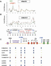
Loss of H4 Arg3 methylation leads to formation of repressive heterochromatin. Results of ChIP with antibodies specific to dimethyl Lys9 (A) and trimethyl Lys27 (B) with mono- and dinucleosomes purified from wild-type (Green) or PRMT1 knock-down (orange) 6C2 cell lines across the β-globin locus. (C) A summary of the ChIP data across the chicken β-globin locus showing results of PRMT1 knock-down. The arrows pointing up and down indicate increase or decrease of modifications, respectively. The minus signs indicate that there are no changes in histone modifications.
Figure 4.
Figure 4.
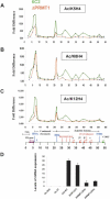
Histone H4 Arg3 methylation is required for histone H4 acetylation at Lys5 and Lys12. (AC) Results of ChIP with antibodies specific to H4 acetylation at Lys5, Lys8, and Lys12 on mono- and dinucleosomes purified from wild-type (green) or PRMT1 knock-down (orange) 6C2 cell lines across the β-globin locus. (D) Quantitative RT–PCR of FR mRNA expression in wild-type (WT), USF1 knock-down, and PRMT1 knock-down (2G5 and 2H2) cell lines.
Figure 5.
Figure 5.
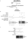
Restoration of histone H4 Arg3 methylation potentiates histone acetylation in vitro. (A) Outline of experimental procedure. (B) The purified oligonucleosomes from wild-type parental 6C2 and siRNA knock-down 6C2 cells were incubated without (left) or with (right) purified PRMT1 in the presence of S-adenosyl methionine (SAM) for 1 h at 30°C. After methylation, the nuclear extracts purified from the same cells and [14C]acetyl-CoA were added and incubated for another 1 h at 30°C as shown. The reaction mixtures were then resolved on SDS-PAGE and visualized by autoradiography. (C) The purified oligonucleosomes from siRNA knock-down 6C2 cells were incubated with purified PRMT1 in the presence or absence of S-adenosyl methionine (SAM) for 1 h at 30°C. After methylation, the nuclear extracts purified from the same cells and [14C]acetyl-CoA were added and incubated for another 1 h at 30°C. The PRMT1 inhibitor AMI-1 (200 μM) was added either before methylation or after methylation.

References

    1. Agalioti T., Chen, G., and Thanos, D. 2002. Deciphering the transcriptional histone acetylation code for a human gene. Cell 111: 381–392. - PubMed
    1. An W., Kim, J., and Roeder, R.G. 2004. Ordered cooperative functions of PRMT1, p300, and CARM1 in transcriptional activation by p53. Cell 117: 735–748. - PubMed
    1. Bannister A.J., Zegerman, P., Partridge, J.F., Miska, E.A., Thomas, J.O., Allshire, R.C., and Kouzarides, T. 2001. Selective recognition of methylated lysine 9 on histone H3 by the HP1 chromo domain. Nature 410: 120–124. - PubMed
    1. Bauer U.M., Daujat, S., Nielsen, S.J., Nightingale, K., and Kouzarides, T. 2002. Methylation at arginine 17 of histone H3 linked to gene activation. EMBO Rep. 3: 39–44. - PMC - PubMed
    1. Bell A.C., West, A.G., and Felsenfeld, G. 1999. The protein CTCF is required for the enhancer blocking activity of vertebrate insulators. Cell 98: 387–396. - PubMed

LinkOut - more resources