. 2005 Aug 15;170(4):559-70.
doi: 10.1083/jcb.200504131.

Sgk3 links growth factor signaling to maintenance of progenitor cells in the hair follicle

Affiliations

Sgk3 links growth factor signaling to maintenance of progenitor cells in the hair follicle

Laura Alonso et al. J Cell Biol. .

Abstract

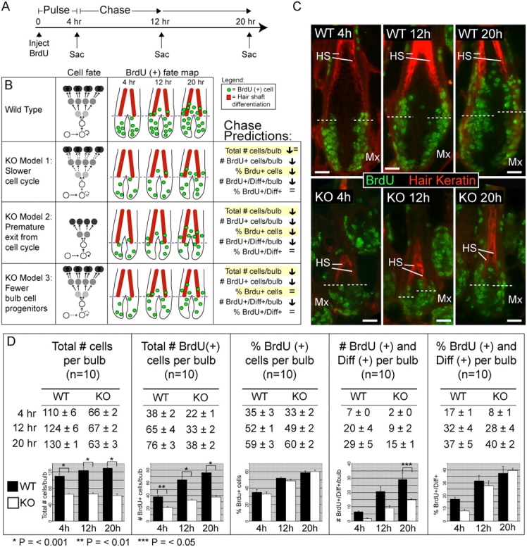
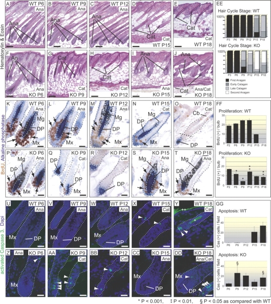
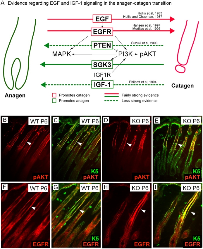
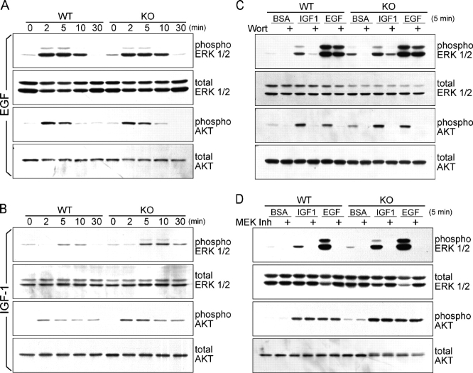
Tyrosine kinase growth factor receptor signaling influences proliferation, survival, and apoptosis. Hair follicles undergo cycles of proliferation and apoptotic regression, offering an excellent paradigm to study how this transition is governed. Several factors are known to affect the hair cycle, but it remains a mystery whether Akt kinases that are downstream of growth factor signaling impact this equilibrium. We now show that an Akt relative, Sgk (serum and glucocorticoid responsive kinase) 3, plays a critical role in this process. Hair follicles of mice lacking Sgk3 fail to mature normally. Proliferation is reduced, apoptosis is increased, and follicles prematurely regress. Maintenance of the pool of transiently amplifying matrix cells is impaired. Intriguingly, loss of Sgk3 resembles the gain of function of epidermal growth factor signaling. Using cultured primary keratinocytes, we find that Sgk3 functions by negatively regulating phosphatidylinositol 3 kinase signaling. Our results reveal a novel and important function for Sgk3 in controlling life and death in the hair follicle.


Figures

Figure 1.
Figure 1.
Targeting of the Sgk3 gene by homologous recombination. (A) Schematic of WT mouse Sgk3 domain structure, gene locus, targeting construct, and mutated Sgk3 allele. Exons 8 and 9 were replaced with PGKneo, and diphtheria toxin A was used for negative selection. Indicated are the antibody binding site, the 5′ external probe A that was used for Southern hybridization, predicted sizes of hybridizing fragments, and primer pairs that were used for PCR. Short arrows represent the primers that were used for PCR analysis of WT (a and b) or mutant (c and d) alleles. RV, EcoRV; Xb, XbaI; S, SmaI. (B) Southern hybridization analysis (SB) of F2 offspring of intercrosses of Sgk3 mutant F1 mice. Tail genomic DNA digested with XbaI was probed with external probe A; W, WT allele; M, mutant allele. (C) Northern analysis (NB) of Sgk3 mRNA expression in total mRNA extracted from P5 skin. EtBr, ethidium bromide. (D) Western immunoblot (WB) shows the presence of Sgk3 in cultured keratinocytes. (E) Immunoblot of dispase-separated epidermis and dermis hair follicle (HF) fractions shows Sgk3 (arrows) in the hair follicle fraction but not in the interfollicular epidermal fraction and also demonstrates that Sgk3 is expressed throughout the hair cycle in whole skin. Extra bands on Western blots are a result of nonspecific signal. Actin, loading control.
Figure 2.
Figure 2.
Sgk3 is required for normal hair coat production. Adult mice (P36 and P411) have a sparse, uneven hair coat (A–D) and irregular hair shafts when seen in profile (E and F). Whiskers are malformed (G and H). Plucked hairs (I and J) lack normal guard, awl, or zigzag hairs. Scanning EM (K and L) shows KO hairs to be irregular and thin with occasional malformed cuticles (arrowheads). Sgk3-null plucked hairs are shorter than WT hairs, with the exception of guard hairs (M). Histology of adult back skin demonstrates that a sparse coat is not caused by the loss of hair follicles (arrows; N–Q). Transmission EM (TEM) of P3 follicles verifies the presence of all cell types in KO follicles (R and S), including the three layers of the IRS. Note the normal keratinization of Henley's layer of the IRS in the upper bulb (asterisks). HS, hair shaft; Ci, IRS cuticle; Hu, Huxley's layer of the IRS; He, Henley's layer of the IRS; ORS, outer root sheath.
Figure 3.
Figure 3.
Hair follicle morphogenesis is normal in Sgk3-null mice until P2. At P2, follicles have normal histology by hematoxylin and eosin (A and B), including ORS, IRS, and hair shaft (HS) layers. Follicle number and density is normal (C and D). Differentiation markers AE13 (hair keratins; hair shaft cortex), AE15 (trichohyalin; IRS), and keratin 6 (K6; companion cell layer [CCL]) are normal in KO follicles at P2 (E–H). Wnt pathway member Lef1 (lymphoid enhancer factor 1) is normal (E and F); mating Sgk3-null mice to Wnt reporter TOPgal mice reveals normal intensity of reporter activity in KO hair follicles at P2 and P6 (I–L). Arrows represent blue Xgal precipitate, demonstrating activity of the TOPgal Wnt reporter. Immunoblot of whole skin lysates at P2 confirms equal amounts of β-galactosidase (M). Mx, matrix; DP, dermal papilla; Epi, epidermis; De, dermis; Pc, precortex; Mg, melanin granules. DAPI, nuclear counterstain; Xgal, β-galactosidase enzymatic substrate. Bars (A, B, and E–H), 20 μM; (C, D, and I–L) 100 μM.
Figure 4.
Figure 4.
Anagen maturation is impaired in Sgk3-null hair follicles. Histology is similar at P2 (A and B) but by P6, Sgk3-null follicles are smaller and fail to grow down to the bottom of the subcutis (C and D). At P6, the shortened KO bulb has a smaller, rounded DP relative to the long, thin DP of WT follicles (G and H). Apoptotic cells are seen in some KO follicle bulbs (J and K); although many P6 bulbs show no apoptotic cells, some have as many as six. Incorporation of BrdU in KO bulbs at P6 is reduced relative to WT bulbs (L–O). Arrows represent examples of BrdU-positive nuclei. (I) Quantification shows that total cell number is reduced in KO bulbs at P6, apoptotic cell number is increased, and proliferation is markedly decreased. Epi, epidermis; De, dermis; SubQ, subcutis; Mx, matrix; Mg, melanin granules; TEM, transmission EM; AP, alkaline phosphatase. Bars (A–D), 100 μM; (E–H and L–O) 20 μM; (J and K) 10 μM.
Figure 5.
Figure 5.
BrdU fate mapping distinguishes between models for reduced bulb cell number and proliferation. (A) Experimental design; mice pulsed with BrdU at P4 were chased for 12 or 20 h. (B) Predicted outcomes for three models. The percentage of Brdu+ cells reflects the percentage of total bulb cells that are BrdU+. The No. of BrdU+/Diff+/bulb reflects the cells per bulb that are positive for BrdU and are above the boundary (dotted lines) of differentiation marker expression. The percentage pf BrdU+/Diff+ is the percentage of BrdU+ cells that are above the boundary relative to the total. Distinguishing between an intrinsic proliferative defect in matrix cells (models 1 and 2) and a defect in the recruitment or regeneration of matrix cells (model 3) relies on the measurement of the percentage of BrdU+ cells. (C) Representative sections demonstrate movement of BrdU-labeled cells. Note that the total number of BrdU-positive cells at 20 h is double the number at 4 h, which is consistent with the expected division after S phase. This confirms that ongoing labeling beyond 4 h does not occur. Dotted lines mark the lower limit of hair keratin expression. HS, hair shaft; Mx, matrix. Bars, 20 μm. (D) Quantification reveals that the most likely explanation for reduced cell number is impaired recruitment or regeneration of progenitors. Error bars represent SEM.
Figure 6.
Figure 6.
Hair follicles lacking Sgk3 enter catagen prematurely. Histology shows the onset of catagen in WT skin at P15 and in KO skin at P9 (A–J). (EE) Quantification of hair cycle stage based on DP morphology that was visualized by alkaline phosphatase (Muller-Rover et al., 2001). Anagen (alI stages); early catagen (catagen I–III); late catagen (IV–VIII). Note that Sgk3-null follicles enter catagen prematurely; by P15, they have entered the second anagen. (K–T) BrdU staining and DP morphology suggest that KO catagen traverses normal stages. Arrows represent BrdU-positive nuclei. (O) Straight dotted line demarcates the bottom of the permanent portion of the hair follicle. (FF) Quantification of BrdU incorporation confirms the appropriate decline in proliferation as KO follicles enter catagen. (U–DD) Staining for activated caspase 3 shows that apoptosis in Sgk3-null follicles increases normally during catagen, excluding massive apoptosis as a mechanism to explain reduced bulb cell number or premature catagen. (Y) Plus signs indicate autofluorescence of the hair shaft (+) and differentiated epidermis (++). Arrowheads, caspase (+) cells. (GG) Quantification of apoptosis confirms the increased number of apoptotic cells in KO skin. Mx, matrix; Ana, anagen; Cat, catagen; Mg, melanin granules; DP, dermal papilla; Cb, Club hair. Error bars represent SEM. Bars (A–J), 100 μM; (K–DD) 20 μM.
Figure 7.
Figure 7.
Growth factor signaling elements are present in the hair follicle. (A) Schematic depicting EGF and IGF-1 pathways and known effects on the anagen to catagen transition. Note the strong evidence that EGF and EGFR promote catagen, which are consistent with the observation that Pten, which is inhibitory to PI3K, may promote anagen. (B–I) Localization of growth factor signaling in the hair follicle. Phospho-Akt is present strongly in the upper ORS, colocalizing with K5, in both WT and KO skin at P6 (B–E). EGFR is expressed in the ORS, colocalizing with K5, and in the interfollicular epidermis and hair bulb (F–I). Arrowheads indicate colocalization with K5 in the ORS.
Figure 8.
Figure 8.
Growth factor signaling is abnormal in Sgk3-null keratinocytes. (A) Treating WT and KO primary keratinocytes with 10 ng/ml EGF reveals normal kinetics of the activation of ERK 1/2 and Akt. (B) In response to 50 ng/ml IGF-1, Sgk3-null keratinocytes exhibit increased activation of ERK 1/2 and Akt. (C) In primary keratinocytes, IGF-1–mediated activation of ERK 1/2 is dependent on PI3K activation, whereas EGF-mediated ERK activation proceeds independently of PI3K. Cells were pretreated with 100 nM wortmannin for 30 min before stimulation. (D) PI3K-dependent IGF-1 activation of ERK does not indicate codependence of PI3K and ERK pathways; phosphorylation of Akt is unaffected by treatment with MEK inhibitor 444937. Cells were pretreated with 1 μM 444937 for 30 min before stimulation. Antibodies are against phosphorylated (activated) or total ERK 1/2 or AKT 1–3 proteins.

Similar articles

Cited by

References

    1. Blanpain, C., W.E. Lowry, A. Geoghegan, L. Polak, and E. Fuchs. 2004. Self-renewal, multipotency, and the existence of two cell populations within an epithelial stem cell niche. Cell. 118:635–648. - PubMed
    1. Bol, D.K., K. Kiguchi, I. Gimenez-Conti, T. Rupp, and J. DiGiovanni. 1997. Overexpression of insulin-like growth factor-1 induces hyperplasia, dermal abnormalities, and spontaneous tumor formation in transgenic mice. Oncogene. 14:1725–1734. - PubMed
    1. DasGupta, R., and E. Fuchs. 1999. Multiple roles for activated LEF/TCF transcription complexes during hair follicle development and differentiation. Development. 126:4557–4568. - PubMed
    1. Datta, S.R., A. Brunet, and M.E. Greenberg. 1999. Cellular survival: a play in three Akts. Genes Dev. 13:2905–2927. - PubMed
    1. Di-Poi, N., C.Y. Ng, N.S. Tan, Z. Yang, B.A. Hemmings, B. Desvergne, L. Michalik, and W. Wahli. 2005. Epithelium-mesenchyme interactions control the activity of peroxisome proliferator-activated receptor beta/delta during hair follicle development. Mol. Cell. Biol. 25:1696–1712. - PMC - PubMed

Publication types

MeSH terms